
Autoimmune hypothyroidism is the end result of an immune attack on the thyroid gland, and the mechanism explains why the gland fails slowly, over years, rather than suddenly.
What the gland looks like in Hashimoto’s thyroiditis
In Hashimoto’s thyroiditis the gland is infiltrated by lymphocytes, and germinal centers form inside it, as they would in a lymph node. This immune tissue replaces the working gland and produces a characteristic picture:
- destruction of follicular cells, most but not all of them
- reduced colloid, the stored hormone-rich material inside the follicles
- mild to moderate fibrosis of the gland
Hashimoto’s thyroiditis and atrophic thyroiditis
Atrophic thyroiditis is the same process at a later stage, and the difference between the two lies in how far the destruction has gone:
- less pronounced infiltration of lymphocytes
- extensive fibrosis
- thyroid follicles that are almost completely absent
Atrophic thyroiditis is therefore the end stage of Hashimoto’s thyroiditis rather than a separate clinical entity. In some cases it has a peculiar fibrosis with a high number of IgG-4 positive plasma cells inside the gland, a variant worth recognizing because it marks the tissue as part of the same autoimmune spectrum.
How the follicular cells die
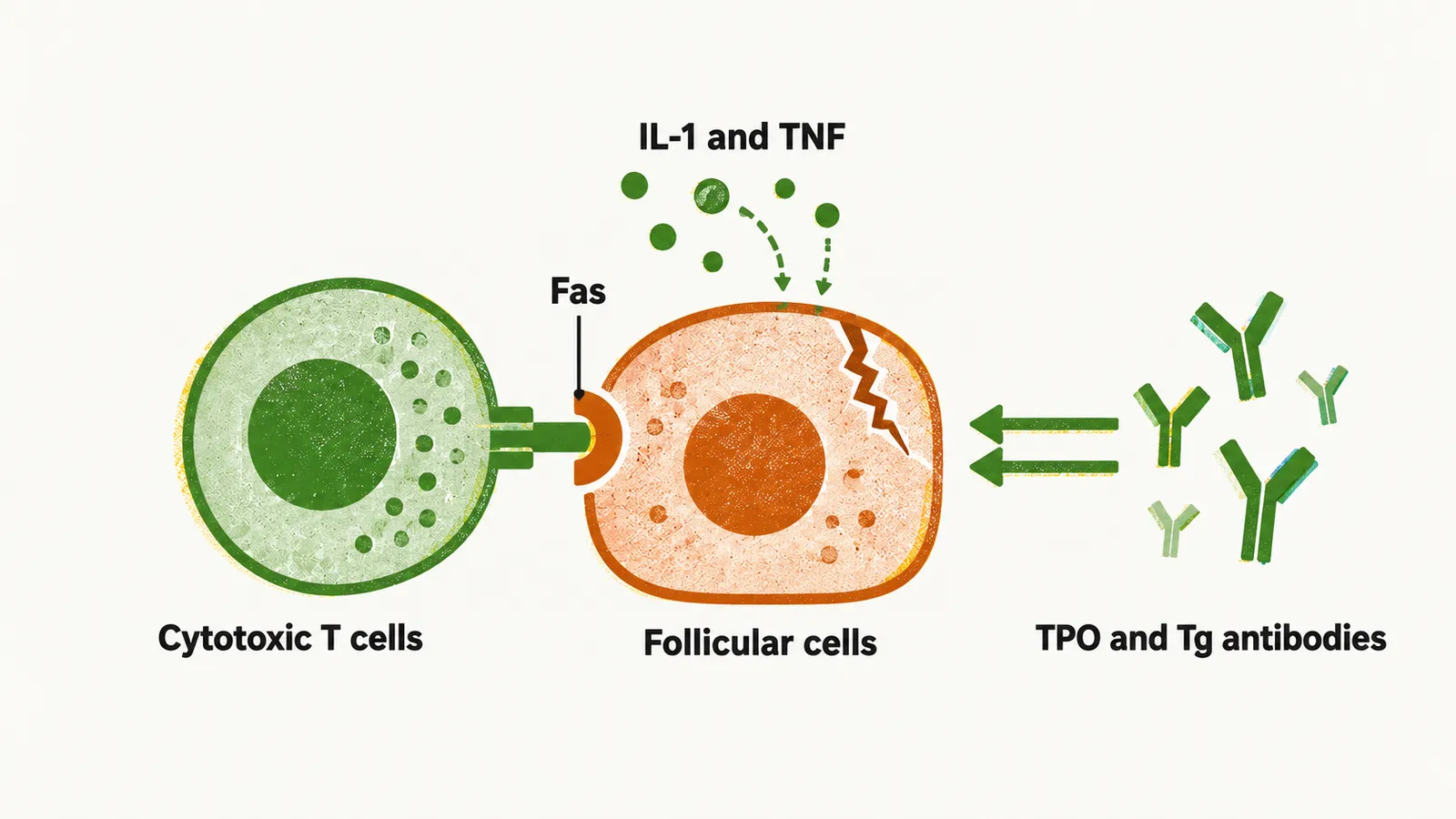
The death of the follicular cells is caused mainly by cytotoxic T cells, lymphocytes that kill other cells directly, and to a minor extent by inflammatory cytokines (signalling molecules of the immune system) such as IL-1 and TNF, which kill the cells through the activation of death receptors such as Fas.
The antibodies against TPO (thyroid peroxidase) and Tg (thyroglobulin) amplify this destruction but do not initiate it. The evidence is what happens in the fetus: maternal TPO and Tg antibodies cross the placenta, yet fetal thyroid destruction does not occur, because the destruction always has to be started by cell-mediated killing. The antibodies are therefore markers and accelerators of the process rather than its trigger.
What raises the risk
The risk of autoimmune hypothyroidism depends on both genetic and environmental factors. The main genetic features that predispose an individual are polymorphisms (common variants) in two genes:
- HLA-DR, which is involved in presenting antigens to T cells
- CTLA-4, a receptor that restrains T-cell activation
HLA-DR and CTLA-4 variants are among the strongest and most widely shared genetic risk factors across autoimmune diseases. This sharing is why patients who already have another autoimmune disease — type 1 diabetes mellitus, Addison’s disease, pernicious anemia, or vitiligo — are more susceptible to autoimmune disease of the thyroid gland as well.
Environmental factors act on this background. Studies show that high exposure to iodine, low exposure to selenium, and low exposure to microorganisms during childhood all increase the chance of autoimmune hypothyroidism. Smoking and alcohol behave in opposite directions: smoking cessation increases the incidence of autoimmune thyroiditis, while people who drink more alcohol have a lower incidence. The finding is an association rather than a recommendation, since the harms of continuing to smoke or drink outweigh any protective signal.
Some drugs, an environmental exposure of a different kind, raise the risk by disturbing regulatory T cells (Treg), the cells whose normal job is to restrain autoimmunity. These include:
- tyrosine kinase inhibitors
- immune checkpoint inhibitors
- alemtuzumab
Because the gland fails slowly, the hormone level falls gradually, and how the disease looks in a patient depends on how far the destruction has gone.