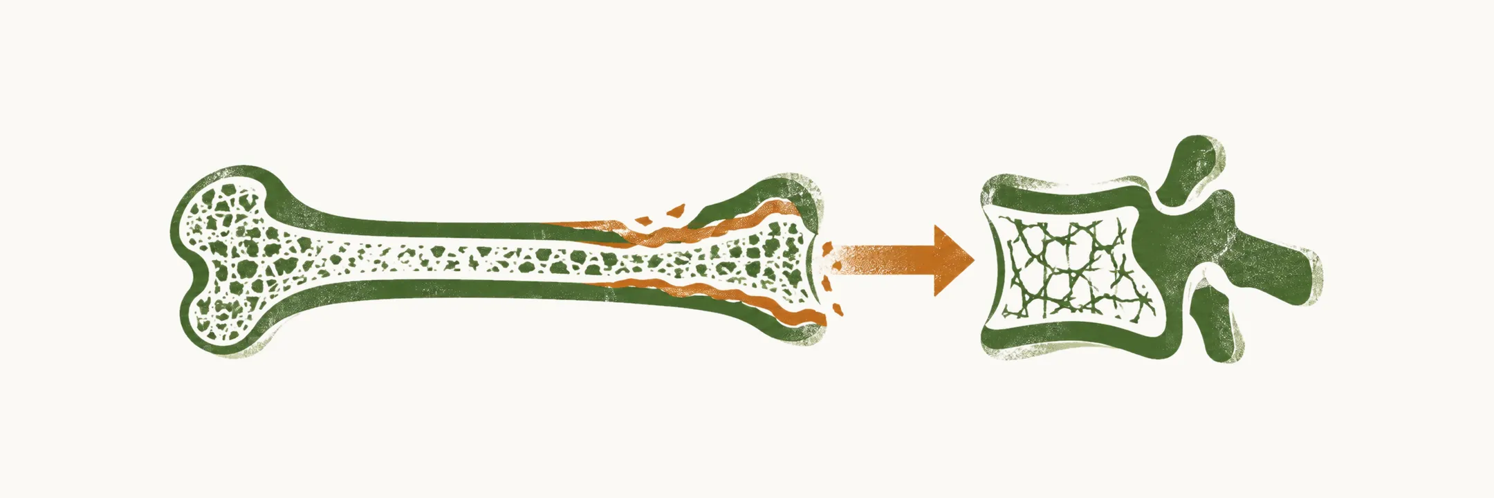
The skeleton is one of the main target organs of parathyroid hormone (PTH), and in primary hyperparathyroidism (PHPT) the excess hormone raises bone turnover. The net loss falls more heavily on cortical bone, the dense outer shell of the long bones, than on trabecular bone, the spongy network inside the vertebrae and the ends of long bones. That single asymmetry explains why the bone disease of PHPT is patchy, why bone density reads differently at different sites, and why fractures cluster where they do.
Skeletal changes
In the past the incidence of skeletal problems caused by PHPT was high, but it is nowadays less than 2% in the US. The classic changes are osteitis fibrosa cystica, a salt-and-pepper appearance of the skull, tapering of the distal third of the clavicle, sub-periosteal bone resorption of the phalanges, and brown tumors.
Osteitis fibrosa cystica is the classic severe picture: resorbed bone is replaced by fibrous tissue, and the brown tumors are the osteoclast-rich fibrous lesions that form within it. These radiographic signs are now rare, but recent advances in bone imaging show that most patients have bone involvement in PHPT even when it is subclinical and asymptomatic.
Bone turnover markers
The markers that can be used to monitor bone turnover separate into those reflecting bone formation and those reflecting bone resorption.
| Bone forming markers | Bone resorption markers |
|---|---|
| Bone-specific alkaline phosphatase | Collagen breakdown products: hydroxyproline, and the pyridinium cross links PYD and DPD |
| Osteocalcin | TRAP, or tartrate-resistant acid phosphatase |
| Amino terminal propeptide of type 1 collagen, or P1NP | NTX and CTX, or the N- and C-telopeptides of type 1 collagen |
Following parathyroidectomy the markers of bone resorption and formation both decrease, but the formation markers decrease more gradually.
Bone densitometry
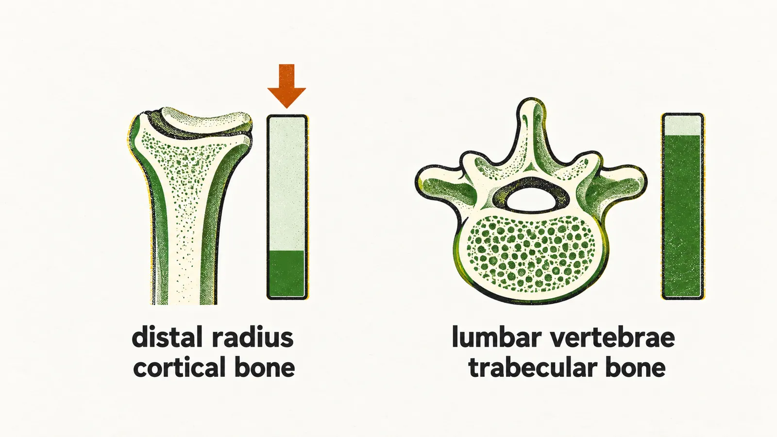
Bone densitometry measures bone mineral density (BMD); the conventional method is dual-energy X-ray absorptiometry (DXA). The density of the bone in patients with PHPT is decreased mainly in cortical bones and not in cancellous (trabecular) bones; therefore, sites with cortical bones are considered the best target for bone densitometry. The site commonly preferred, because it has enough cortical bone and is easy to reach, is the distal third of the radius. In patients with PHPT there is a reduction of bone density at the distal third of the radius, but minimal change in the lumbar vertebrae.
Bone histomorphometry
Bone histomorphometry measures bone turnover directly, on histological study of a bone biopsy taken mainly from the iliac crest. It can measure both static and dynamic changes: static means features of the bone at one time point, and dynamic means comparing two or more snapshots in time. To measure the dynamic values the patient is given dyes, and the change in the location of that dye, driven by the balance of bone formation and bone resorption, is then read.
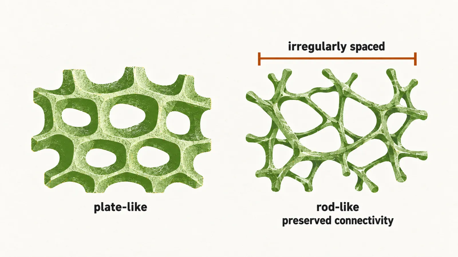
One of the most important indices it measures is trabecular density, and also trabecular connectivity, the degree to which the trabecular struts remain joined to one another. Trabecular connectivity in patients with PHPT is preserved, because PTH acts more on cortical bone than on trabecular bone. Trabecular connectivity is reduced in the iliac crest in postmenopausal women with estrogen-dependent osteoporosis, and in fact patients with PHPT have higher trabecular density and connectivity in comparison with a healthy control group.
Preserved or even increased trabecular density and connectivity does not mean that the trabecular bone is healthy. In these patients, even during the early stages and in a mild condition, there is pathology in the trabecular bones as well:
- thinning of trabecular bone
- more irregularly spaced trabeculae
- rod-like rather than plate-like organization
So a paradox arises: the bone mineral density in patients with PHPT is preserved in the vertebrae, yet at the same time there is an increase of vertebral fracture. Understanding it requires the newer imaging techniques.
High-resolution peripheral quantitative computed tomography (HRpQCT)
In HRpQCT the 3D density is measured, not the 2D plate-like density that is measured by conventional DXA, so it is more precise. HRpQCT is mainly used to measure bone density in the radius and the tibia. Studies with HRpQCT show that patients with PHPT have a decrease of bone trabecular density, which opposes what is seen with histomorphometry. Together the two findings answer the paradox of the increase of vertebral fracture despite a vertebral BMD that DXA shows as preserved: trabecular bone connectivity is preserved or even increased, but the quality of the bone is very low and the trabeculae are thin, which makes them prone to fracture.
Because cortical and trabecular bone are affected differently, bone fracture in patients with PHPT can be described as highly site-specific.