
Diagnosing primary hyperparathyroidism (PHPT) means showing hypercalcemia together with a high, or inappropriately normal, parathyroid hormone (PTH) level, and then excluding the other causes of that picture.
Diagnosis
The diagnosis of PHPT is done based on two grounds:
- high serum calcium level plus high PTH (usually within 2 times above the upper limit of normal, ULN)
- high serum calcium level plus inappropriately normal PTH
The phrase “inappropriately normal or elevated” needs a precise meaning. Physiologically, in hypercalcemia the PTH is suppressed to the lower limits of normality or even lower; therefore, in a patient with hypercalcemia, even if the value of PTH is in the normal range, 10-65 pg/ml, it is not normal.
The normal range for PTH is quite wide, between 10-65 pg/ml, which is reflective of physiological change that happens due to:
- age, by which the PTH level will increase within the normal range
- vitamin D level, by which the normal range will be extended in populations that are vitamin D deficient
- renal function, since patients with renal dysfunction tend to have higher levels of PTH due to abnormal clearance and degradation of PTH
Other biochemical changes
Besides calcium and PTH, PHPT changes other laboratory values:
- low phosphate level, but within the normal range; frank hypophosphatemia happens in only 25% of the cases
- hypercalciuria in 40% of the cases
- 25-hydroxy vitamin D in the low-normal range, and 1,25-dihydroxy vitamin D in the upper-normal range, or increased, in 30% of the cases; both are caused by the effect of PTH on the 1α-hydroxylase of the renal tubules, which converts 25-hydroxy vitamin D to calcitriol (1,25-dihydroxy vitamin D)
The values below are the mean ± SEM in a group of patients with PHPT, set beside the normal ranges:
| Parameter | Patient values (mean ± SEM) | Normal range |
|---|---|---|
| Serum calcium | 10.7 ± 0.1 mg/dL | 8.2-10.2 mg/dL |
| Serum phosphorus | 2.8 ± 0.1 mg/dL | 2.5-4.5 mg/dL |
| Total alkaline phosphatase | 114 ± 5 IU/L | <100 IU/L |
| Serum magnesium | 2.0 ± 0.1 mg/dL | 1.8-2.4 mg/dL |
| PTH (IRMA) | 119 ± 7 pg/mL | 10-65 pg/mL |
| 25(OH)D | 19 ± 1 ng/mL | 30-80 ng/mL |
| 1,25(OH)2D | 54 ± 2 pg/mL | 15-60 pg/mL |
| Urinary calcium | 240 ± 11 mg/g creatinine | - |
| Urine DPD | 17.6 ± 1.3 nmol/mmol creatinine | <14.6 nmol/mmol creatinine |
| Urine PYD | 46.8 ± 2.7 nmol/mmol creatinine | <51.8 nmol/mmol creatinine |
Differential diagnosis
Once hypercalcemia with a high or inappropriately normal PTH is found, the other forms of hyperparathyroidism and the other causes of hypercalcemia have to be separated from PHPT. The table compares the biochemical pattern of each.
| Cause | Serum calcium | PTH | Urine calcium |
|---|---|---|---|
| Primary hyperparathyroidism | Elevated | Elevated or inappropriately normal | High or high normal |
| Hypercalcemia of malignancy and non-PTH mediated hypercalcemia | Elevated | Suppressed | Typically high |
| Secondary hyperparathyroidism | Normal or low | Elevated | Low in vitamin D deficiency, malabsorption, chronic renal failure; high in idiopathic hypercalciuria |
| Tertiary hyperparathyroidism | Elevated | Elevated | Low before transplant |
| FHH | Elevated | Typically high normal or elevated | Typically low |
| Normocalcemic primary hyperparathyroidism | Normal | High | <350 mg/24 hours |
Other forms of hyperparathyroidism
- secondary hyperparathyroidism: characterised by a high level of PTH and hypocalcemia, which mostly happens in vitamin D deficiency; other causes can be renal failure and malabsorption.
- tertiary hyperparathyroidism: happens in patients with severe and prolonged secondary hyperparathyroidism, such as renal transplant patients with a history of dialysis. In these cases there is formation of an autonomous hyperfunctioning adenoma in the parathyroid gland after a long period of hypocalcemia.
Hypercalcemia of malignancy and other causes of hypercalcemia
One of the most important steps in the differential diagnosis of PHPT is hypercalcemia of malignancy.
Usually, in malignant patients there are:
- symptomatic patients
- severe hypercalcemia
- decreased serum PTH
In some cases of malignancy-related hypercalcemia there is an increase of production of PTHrP, or PTH-related protein, which cross-reacts with the PTHR-1 receptor and leads to hypercalcemia.
While more than 90% of the cases of hypercalcemia are related to either PHPT or malignancy, the other possible causes of hypercalcemia for differential diagnosis are:
- vitamin D intoxication
- granulomatous disease
- familial hypocalciuric hypercalcemia, or FHH
- lithium use
- thiazide use
In patients with a history of lithium and thiazide use, if withdrawing these drugs corrects the hypercalcemia and the high PTH level within 3-6 months, then the hypercalcemia was caused by the drug; if it does not, PHPT is confirmed.
In patients with FHH there is mostly a mutation in the gene of the CaSR, which leads to hypercalcemia together with a low concentration of calcium in the urine. FHH is present from birth, so a patient whose hypercalcemia is first discovered after the age of 50 is unlikely to have it, and a urinary calcium to creatinine clearance ratio below 0.01 supports FHH over PHPT.
Normocalcemic PHPT
In a normocalcemic patient with a high PTH, normocalcemic PHPT, or NPHPT, can be confirmed only if all the causes of secondary hyperparathyroidism are excluded. In fact, the diagnosis of NPHPT has a high occurrence of mis-diagnosis; for instance, in a patient with PHPT and concomitant vitamin D deficiency there will be a balance between the increase of calcium caused by PTH and the decrease of calcium caused by the vitamin D deficiency. Here the diagnosis is PHPT with vitamin D deficiency, not NPHPT; therefore, the diagnosis of NPHPT should always be made carefully.
Diagnostic workup
For the diagnosis of PHPT it is suggested to perform:
Laboratory tests
- PTH level
- serum calcium level: both the adjusted and ionized form
- serum phosphate level
- 24-hour urinary calcium
- vitamin D level, for determining the hypocalcemic effect of vitamin D deficiency
- alkaline phosphatase
- serum electrophoresis: albumin level
- creatinine, for evaluation of kidney function
Imaging
- DEXA (dual-energy X-ray absorptiometry)
- upper abdomen ultrasound, for evaluation of nephrolithiasis
- thoracolumbar X-ray, for evaluation of silent vertebral fractures
Not mandatory for diagnosis:
- neck ultrasound
- scintigraphy
- MRI or CT of neck
Treatment
For symptomatic patients, parathyroidectomy is the first-line treatment, because removing the overactive gland or glands is the only definitive cure and takes away the source of the excess PTH.
For asymptomatic patients with PHPT, surgery is first-line when at least one of the conditions below is met:
- serum calcium more than 1 mg/dl above the ULN
- bone mineral density by DEXA T-score ≤ -2.5, in each of the lumbar spine, total hip, femoral neck, or distal third of the radius
- vertebral fracture on imaging by X-ray, CT, MRI, or vertebral fracture assessment
- creatinine clearance < 60 cc/min
- 24-hour urine calcium > 400 mg/day together with an increased biochemical stone risk profile
- nephrolithiasis or nephrocalcinosis found by ultrasound, CT, or X-ray
- age under 50 years
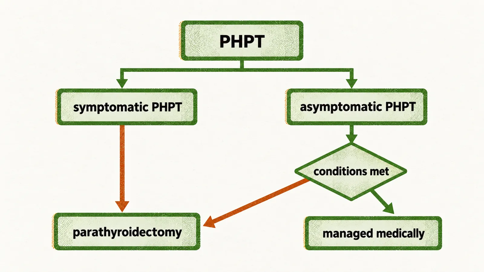
Patients who do not meet any of these conditions, or who cannot have or decline surgery, are managed medically: calcium and vitamin D are kept at normal levels, and where the hypercalcemia or bone loss needs treating, a calcimimetic such as cinacalcet lowers PTH and serum calcium while a bisphosphonate improves bone mineral density without correcting the hypercalcemia.