
Philadelphia-negative myeloproliferative neoplasms (MPNs) are driven by mutations that keep a single intracellular signalling pathway switched on. The clinical picture is not random: it follows the burden of the cells that carry the mutation.
Driver events
The driver events in Philadelphia-negative MPN are mutations in the JAK2, CALR and MPL genes. These three mutations are largely mutually exclusive and all converge on the same pathway, so any one of them is enough to start the disease. In more than half of the cases, other mutations can be found as well, such as DNMT3A, ASXL1 and TET2, genes that normally regulate the epigenetic marks that control how DNA is read.
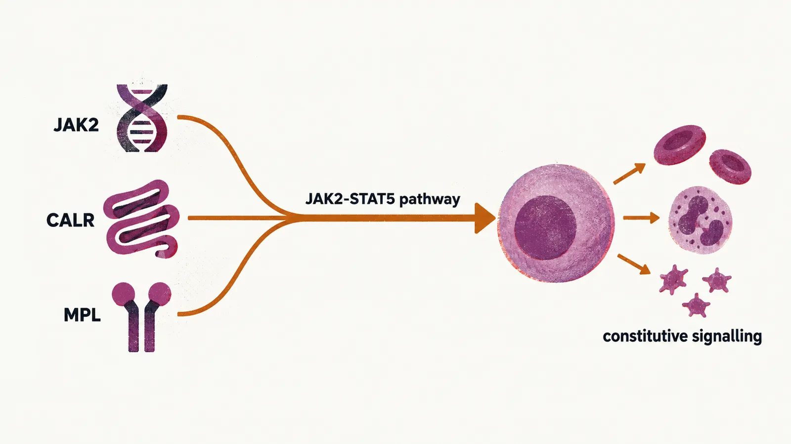
The mutation that is found most often is JAK2-V617F. It affects the part of the JAK2 protein that controls its basal activity, so the protein no longer waits for an external signal before firing, and the result is an increase of uncontrolled activity of the JAK2-STAT5 pathway. That constitutive signalling is what drives the marrow to overproduce mature blood cells.
The natural history of this most prevalent mutation starts with a point mutation in the JAK2 gene, which is located on chromosome 9. Through mitotic recombination, both copies of chromosome 9 in the cell then carry the same genetic defect, so a single-cell clone becomes homozygous for the mutation and the abnormal signal is doubled.
Allele burden and the clinical picture
How much of the pathway is active depends on how much of the marrow carries the mutation, and the clinical picture follows the same gradient. Studies show that the clinical manifestations of all the JAK2-V617F disorders described so far correlate with the burden of JAK2-V617F positive cells — the allele burden — found in the bone marrow or peripheral blood, in the following way:
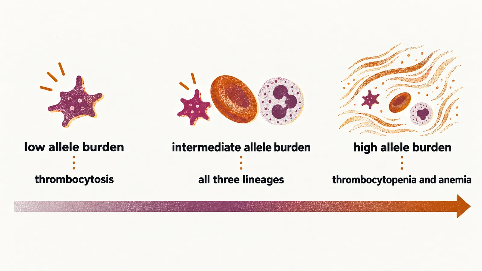
- low allele burden: the only clinical manifestation is mostly thrombocytosis, a raised platelet count
- intermediate allele burden: most of the time all three lineages are involved, which produces thrombocytosis, erythrocytosis (a raised red cell mass) and leukocytosis (a raised white cell count)
- high allele burden: most of the time all three lineages are involved, and perhaps because of the high rate of fibrosis here, there is thrombocytopenia, anemia or erythrocytosis, and leukocytosis
Because the same pathway is switched on in polycythemia vera (PV) and in primary myelofibrosis and post-essential-thrombocythemia myelofibrosis, ruxolitinib, a competitive inhibitor of JAK proteins, is effective in those diseases.
Inflammatory signalling
Beyond its role in the stem cells, where it drives an unwanted production of cells when the JAK2 pathway is switched on, JAK2 is also the intracellular pathway for many inflammatory signals. Studies show that the increase of inflammatory cytokines in these patients carries a prognostic effect: higher circulating cytokine levels are associated with a heavier symptom burden and a worse prognosis. JAK inhibitors suppress these cytokines, which is part of how they relieve the constitutional symptoms of the disease.
Most of the cases of MPNs are diagnosed in the chronic phase, the early stage of the disease; the chance that the disease later progresses to the blastic phase, the stage in which immature blast cells accumulate, reflects the accumulation of additional mutations in the clone over time.