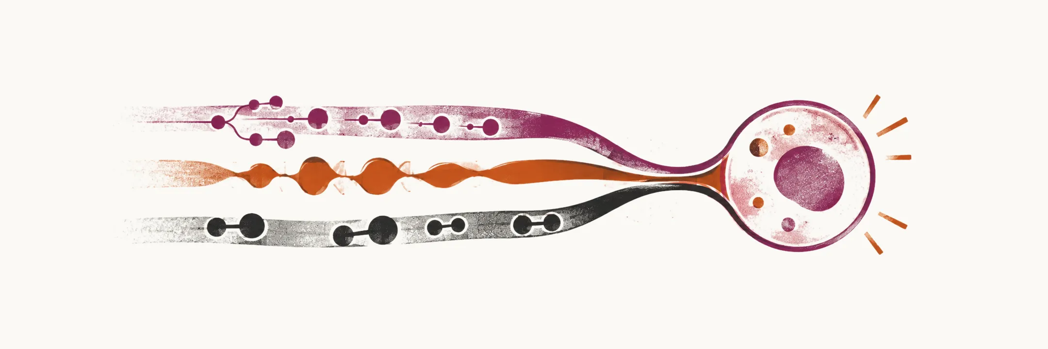
The myeloid neoplasms are clonal diseases of the myeloid lineage, the branch of haematopoiesis that gives rise to granulocytes, monocytes, red blood cells and platelets. Some leave the bone marrow producing too few mature cells, and others too many, and the two behaviours overlap: the same kind of clone can tip either way. Naming the disease precisely matters because the name predicts what a bone marrow biopsy should show, which mutation is worth testing for, and what the treatment will be.
The fifth edition of the WHO classification of haematolymphoid tumours (WHO-HAEM5) defines each entity through a triad of attributes: its lineage, its dominant clinical attribute and its dominant biologic attribute. Lineage is assigned by immunophenotyping, using flow cytometry and/or immunohistochemistry to read the surface and cytoplasmic markers of the cells, because those markers identify the lineage the cells belong to. The dominant clinical attribute describes how the untreated disease behaves, using the general descriptors acute or chronic and cytopenia (myelodysplasia) or cytosis (myeloproliferation). The dominant biologic attribute is the gene fusion, rearrangement or mutation that defines the entity. Asking the same three questions of every disease is what keeps the classification consistent from one entity to the next.
The classification then runs along lineage, broadly from benign to malignant, and branches into category, family, type (the disease or tumour itself) and subtype. It is a framework rather than a flat list, so an entity is understood by the branch on which it sits.
The chronic myeloid neoplasms are one family, in which the myeloproliferative neoplasms sit beside the myelodysplastic neoplasms and the disorders that combine features of both. The relation between pre-neoplastic and neoplastic myeloid disorders is close, because they arise from the same haematopoietic stem cell. That is why the myeloproliferative neoplasms are learned together with the rest of the myeloid neoplasms: the individual diagnoses only make sense once their place in the family is clear.
Within the chronic myeloid neoplasms, several distinct entities are recognised, and they are separated by two questions that are used side by side.
Philadelphia status
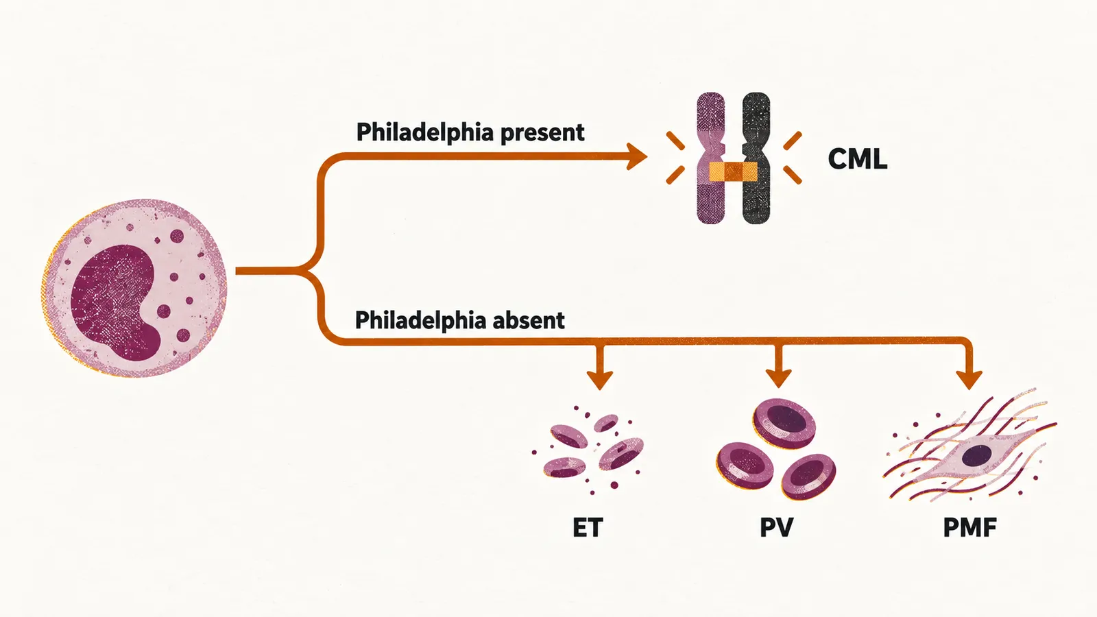
The first question is whether the Philadelphia translocation is present. It creates the BCR::ABL1 fusion and a constitutively active tyrosine kinase, meaning one that is switched on without an external signal, and it is the defining abnormality of chronic myeloid leukaemia (CML) — the only Philadelphia-positive myeloproliferative neoplasm. When the Philadelphia translocation is absent, the classical myeloproliferative neoplasms are mostly driven by JAK2-related mutations, and they fall into three entities:
- essential thrombocythemia (ET)
- primary myelofibrosis (PMF)
- polycythemia vera (PV)
The lineages involved
The second question is which lineages are increased in the peripheral blood, because that is where the different members of the family show themselves first. In CML the proliferation is dominated by the granulocytic lineage, with basophilia (a raised basophil count) and often a raised platelet count. Chronic neutrophilic leukaemia (CNL) and chronic eosinophilic leukaemia (CEL) are much rarer relatives in which a single granulocytic lineage dominates.
In ET, PV and PMF the lineage pattern differs, and it is a clue to which entity is present. PV is a panmyelosis, so all three lineages are increased: the red cells rise, and the platelets and white cells are often raised as well. PMF involves the megakaryocytic and granulocytic lineages and produces marrow fibrosis, and it raises or lowers the counts as that fibrosis develops. ET is mainly a proliferation of the megakaryocytic (platelet) lineage, which is why its main manifestation is thrombocytosis while the marrow keeps essentially normal erythropoiesis and granulopoiesis. All of these are clonal stem-cell disorders, so the lineages that appear raised in the blood are a clue rather than the whole definition; the bone marrow morphology and the driver mutation complete it.