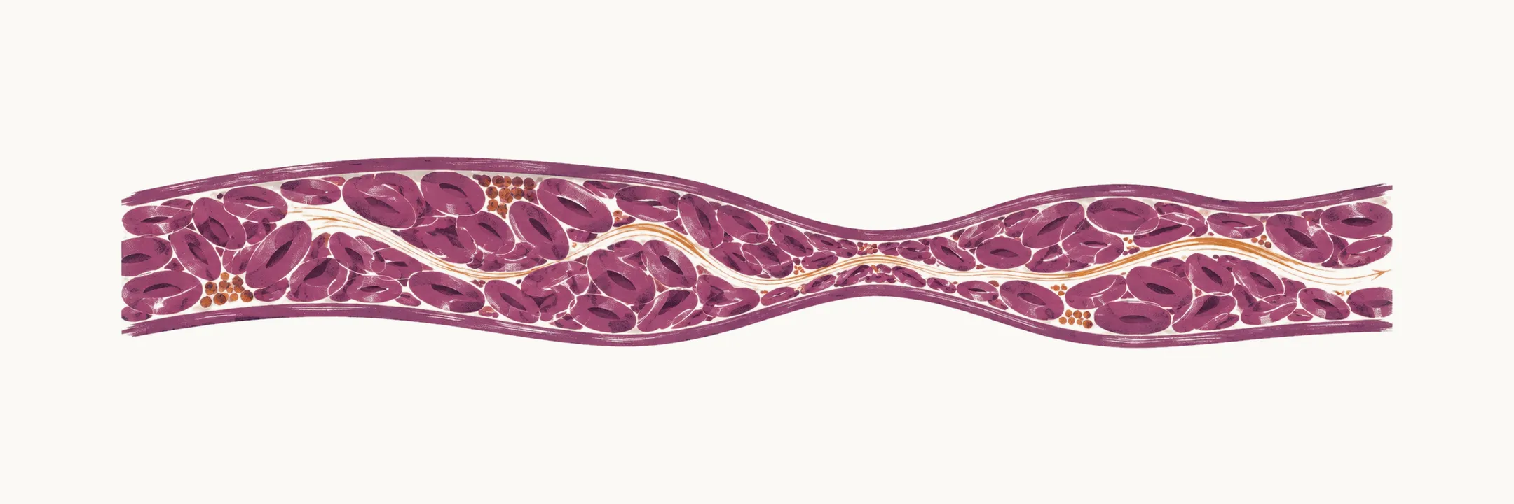
Polycythemia vera (PV) is a myeloproliferative neoplasm in which erythrocytosis, a raised red cell mass, is the most common presentation. Because PV is a panmyelosis, in which all three lineages of myeloid cells are increased, the red cell mass rises and the platelet count is often raised as well; the resulting hyperviscosity and thrombocytosis together make thrombosis the most important complication.
The incidence of PV is between 0.8 and 2.6 cases per 100,000.
Presentation and complications
The main clinical presentations in PV are:
- erythrocytosis
- thrombocytosis
- leukocytosis of different magnitude
The most common one is erythrocytosis, with a burden of 80%, while for thrombocytosis and leukocytosis it is about 40% of cases.
The main signs of PV are related to the massive increase of red blood cell mass, and they are:
- hypertension
- blood hyperviscosity
- thrombotic events
Thrombotic events can be seen in 20% of cases as the initial sign, and the incidence rate of thrombotic events in PV is reported as 3.3% per person per year. The risk factors for vascular events are advanced age, a history of thrombosis and leukocytosis, together with other factors such as obesity, hypertension and smoking. In the case of splanchnic vein thrombosis, PV has to be considered as part of the differential diagnosis.
Myelofibrotic progression is reported in 4.9% to 6% of cases at 10 years (secondary myelofibrosis, SMF). In the case that the patient has post-polycythemia vera myelofibrosis (PPV-MF) or post-essential thrombocythemia myelofibrosis (PET-MF), a prognostic model called the MYSEC prognostic evaluation can be used.
Diagnosis
The diagnosis of PV is based on the presence of:
- high concentration of hemoglobin or high hematocrit
- panmyelosis, which is all three lineages of myeloid cells
- pleomorphic mature megakaryocytes in the bone marrow
- presence of JAK2-related mutations (in 99% of cases)
The criteria of the WHO for the diagnosis of PV, based on the 2022 classification, set the threshold for high hemoglobin or hematocrit as:
- Men: hemoglobin >16.5 g/dl, or hematocrit >49%
- Women: hemoglobin >16 g/dl, or hematocrit >48%
Ruling out secondary erythrocytosis
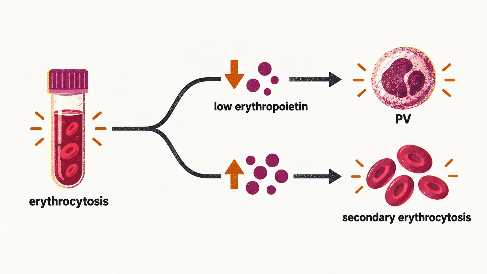
In erythrocytosis, secondary erythrocytosis must always be considered and ruled out; it is mostly seen in isolated erythrocytosis, and it can be due to any cause that produces low oxygen or a high erythropoietin level:
- low oxygen: high altitude, respiratory disorders, cardiac shunt, smoking
- high erythropoietin: renal disorders; uterus, kidney and liver neoplasms
Taking the history of the patient who has erythrocytosis helps to differentiate PV from the other causes; the history should cover drugs such as SGLT2 inhibitors and androgens, smoking habit, erythropoietin level, oxygen level, lung function test, and a scan of the abdomen. Low erythropoietin is predictive of PV, because a genuinely raised red cell mass suppresses erythropoietin, whereas in secondary erythrocytosis the hormone is driving the marrow and is therefore normal or high.
Risk-adapted management
Management is matched to the risk group. Every patient has cardiovascular risk factors managed and takes aspirin 81-100 mg/day, phlebotomy is used to reach a hematocrit below 45%, and high-risk disease adds cytoreductive treatment with hydroxyurea.
Low risk.
- manage cardiovascular risk factors
- aspirin 81-100 mg/day
- phlebotomy to reach a hematocrit of less than 45%
Phlebotomy is used rather than a drug because removing red cells directly lowers the blood viscosity, and the target is a hematocrit below 45%, the level above which the thrombotic risk rises.
Follow-up consists of:
- monitoring for new thrombosis or acquired von Willebrand disease (VWD) and/or any major bleeding problems
- evaluating the patient for any cytoreduction therapy indication
- monitoring signs and symptoms of disease progression
Patients who are asymptomatic and have no indications for cytoreduction therapy continue aspirin and monitoring. A potential indication for cytoreduction therapy, present when the patient is symptomatic, is:
- new thrombosis or acquired VWD
- frequent phlebotomy or intolerance to phlebotomy
- progressive thrombocytosis or leukocytosis
- disease-related symptoms such as night sweats and weight loss
- splenomegaly
Disease progression leads to post-PV myelofibrosis (MF).
High risk.
- manage cardiovascular risk factors
- aspirin 81-100 mg/day
- phlebotomy to reach a hematocrit of less than 45%
- a regimen for cytoreductive treatment, which is hydroxyurea
Follow-up consists of:
- monitoring for new thrombosis or acquired VWD and/or any major bleeding problems
- monitoring signs and symptoms of disease progression
An adequate response is followed by continuing aspirin and monitoring. An inadequate response or a loss of response is a potential indication for a change of cytoreductive therapy:
- intolerance or resistance to the previous agent
- new thrombosis
- frequent phlebotomy
- splenomegaly
- progressive leukocytosis or thrombocytosis
- disease-related symptoms such as night sweats and weight loss
In that case the change of cytoreductive therapy is ruxolitinib. Disease progression leads to post-PV MF or to the accelerated or blastic phase.