
Essential thrombocythemia (ET) is a myeloproliferative neoplasm whose main clinical manifestation is thrombocytosis, a raised platelet count. Its incidence is estimated to be about 0.8 to 1.2 cases per 100,000, with a median age of 58 years and a female predominance of about 2:1.
Diagnosis
The most important thing in the initial evaluation of a patient who is suspected of having ET is to differentiate it from the other causes of thrombocytosis, which are:
- infections
- inflammatory disorders
- solid cancers
- chronic bleeding
These causes raise the platelet count through a reaction to something else, whereas ET raises it through a clone that makes platelets autonomously. More than 50% of cases of ET are asymptomatic and are diagnosed incidentally, with thrombocytosis above 450 × 10^9/L as the primary sign. In many patients the platelet count itself is the only clue, but the main signs of ET in most of the patients are thrombotic or hemorrhagic symptoms.
Mild splenomegaly can be seen in about 10% of cases, while hepatomegaly is not frequent at all. Leukocytosis can also be seen, but it is not that prevalent; it is found in about 20% of cases.
The bone marrow provides the morphological confirmation. Based on the WHO, the morphological changes that can be seen in ET are:
- megakaryocytic proliferation
- mature megakaryocytes
- normal erythropoiesis and granulopoiesis
- normal cellularity of the bone marrow
Thrombotic and hemorrhagic risk
ET carries a risk of both thrombosis and bleeding. The rate of fatal and non-fatal thrombotic events is estimated at about 1.9% per patient per year, and in the natural history of patients who have ET, thrombotic events are seen in 20% of cases.
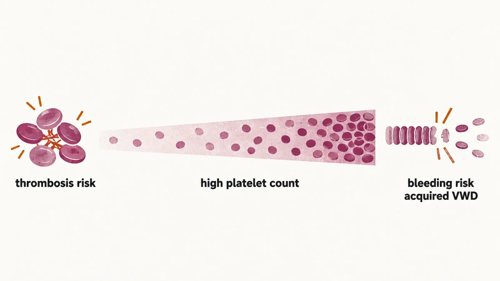
The risk of thrombotic events is increased by advanced age, a history of thrombosis, leukocytosis and the presence of JAK2 mutations. Thrombosis risk in patients with ET can also be estimated with a dedicated model, the IPSET-t model.
At the other end of the spectrum, a very high platelet count can predispose to bleeding. In acquired von Willebrand disease (VWD) the excess platelets remove the largest von Willebrand factor multimers from the plasma, and because those multimers matter most for platelet adhesion, their loss produces a bleeding tendency.
Risk-adapted management
| Risk group | Criteria | Management |
|---|---|---|
| Very low | age < 60 years, no JAK mutation, no prior history of thrombosis | manage cardiovascular risk factors; aspirin 81-100 mg/day for patients who have vasomotor or microvascular disturbances |
| Low | age < 60 years, with JAK mutation, no prior history of thrombosis | manage cardiovascular risk factors; aspirin 81-100 mg/day |
| Intermediate | age > 60 years, no JAK mutation, no prior history of thrombosis | manage cardiovascular risk factors; aspirin 81-100 mg/day |
| High | age > 60 years, with JAK mutation, prior history of thrombosis at any age | manage cardiovascular risk factors; aspirin 81-100 mg/day; cytoreductive therapy, which is hydroxyurea |
For very-low, low and intermediate risk, follow-up consists of:
- monitoring for new thrombosis or acquired von Willebrand disease (VWD) and/or any major bleeding problems
- evaluating the patient for any cytoreduction therapy indication
- monitoring signs and symptoms of disease progression
Patients who are asymptomatic and have no indications for cytoreduction therapy continue aspirin and monitoring. A potential indication for cytoreduction therapy, present when the patient is symptomatic, is:
- new thrombosis or acquired VWD
- splenomegaly
- progressive thrombocytosis or leukocytosis
- disease-related symptoms such as night sweats and weight loss
- vasomotor or microvascular disturbances without response to aspirin, such as headache, chest pain and erythromelalgia
Disease progression leads to post-ET myelofibrosis (MF).
For high risk, follow-up consists of:
- monitoring for new thrombosis or acquired VWD and/or any major bleeding problems
- monitoring the response to treatment
- monitoring signs and symptoms of disease progression every 3-6 months
Prognosis
The median survival of ET patients is about 14 to 21 years. Besides progression to post-ET myelofibrosis, progression to the accelerated phase (AP) or the blastic phase (BP), the later stages in which blast cells increase, is also possible in patients with ET: for the AP in 10 years it is estimated at 0.7-3%.