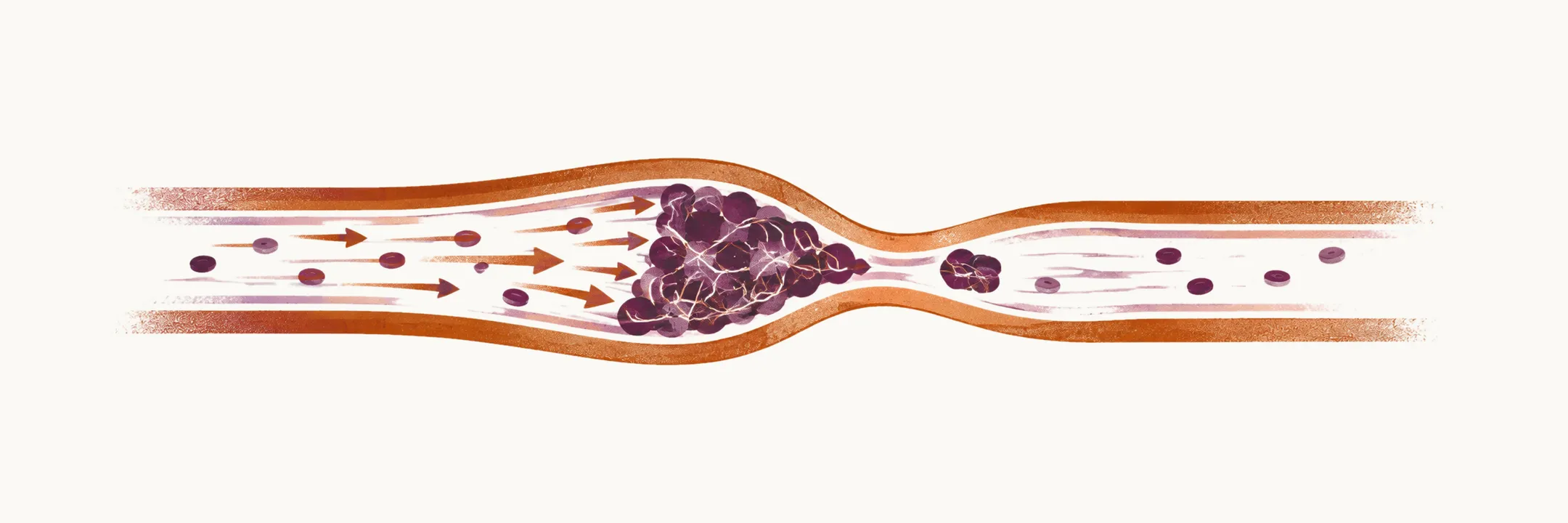
Thrombosis is the obstruction of blood flow due to the formation of a clot, or thrombus. It may result in anoxia (a lack of oxygen in the tissue) and tissue damage, and it is the major reason for morbidity and mortality in venous and arterial diseases. When a thrombus that has formed in a venous or arterial part of the circulation spreads, it is called embolism.
Hemostasis is the process that stops bleeding from a damaged vessel. The factors that control hemostasis are the main factors that control thrombosis, so hemostasis and thrombosis sit in an interplay between three factors:
- vessel walls
- coagulation and fibrinolytic pathways, which form clots and break them down
- platelets
The causes of thrombotic disorders are grouped under Virchow triad: stasis of blood flow, injury to the vessel wall, and a hypercoagulable state, meaning an increased tendency of the blood to clot.
Which of these factors matters most depends on where the thrombus forms, and this is the idea that organises the topic. Thrombosis is classified into two groups based on the anatomical location where it occurs:
- venous thrombosis is dependent on a hypercoagulable state of the body, due to coagulation or fibrinolytic abnormalities, or on stasis of blood flow in an organ
- arterial thrombosis is highly dependent on the blood flow, the vessel wall and platelet abnormalities
Choose a route through the topic
Because the two forms are driven by different factors, they are best studied one after the other. Start with arterial thrombosis: the platelet and vessel wall abnormalities it describes are the ones the platelet pathophysiology note then follows in detail. The venous notes then take up the other form, from its causes through diagnosis, treatment and prevention.
- Arterial thrombosis, its risk factors and antiphospholipid syndrome explains why platelet and vessel wall abnormalities drive thrombosis in the arteries, the risk factors for arterial thrombosis, and antiphospholipid syndrome with its three clinical forms, its mechanism and the laboratory tests used in diagnosis.
- Platelet pathophysiology in arterial thrombosis follows the platelet from its adhesion to the exposed vessel wall through activation and irreversible aggregation, covering its granules, its surface receptors, and the ways inflammation and infection increase platelet reactivity.
- Venous thrombosis, its risk factors and inherited causes covers the acquired risk factors for venous thrombosis and the inherited defects that raise clotting risk, including antithrombin, protein C and protein S deficiency, factor V Leiden and the G20210A variant of factor II, and the contribution of impaired fibrinolysis.
- Diagnosing deep-vein thrombosis and pulmonary embolism gives the incidence and short-term fatality of deep-vein thrombosis and pulmonary embolism, how each presents, and the diagnostic sequence from clinical probability scoring to D-dimer testing, compression ultrasonography and CT pulmonary angiography.
- Screening for thrombophilia and treating venous thromboembolism sets out when testing for inherited thrombophilia is worthwhile, and which anticoagulants are used to treat venous thromboembolism.
- Preventing venous thromboembolism explains how acutely ill patients are assessed for venous thromboembolism risk and which mechanical and pharmacological measures are used to prevent it.