
Deep-vein thrombosis (DVT) is a thrombus in a deep vein, most often of the legs, and pulmonary embolism (PE) is the embolisation of such a thrombus to the lungs. Both are forms of venous thromboembolism (VTE). DVT and PE occur in 1 to 2 cases per 1000 individuals each year in the US.
The 28-day fatality rate from DVT is about 9%, and from PE it is about 15%. PE is the more immediately dangerous: it is the form that can present as sudden death.
How each presents
Neither condition can be recognised reliably from its clinical features alone. Clinical signs of DVT are non-specific and insensitive: many DVTs cause no signs at all, and the signs that do appear also occur in other leg conditions. The main signs of DVT are:
- swelling or edema
- pain
- warmth
PE has no single classic picture. The symptom that appears most often is sudden dyspnea (breathlessness), followed by pleuritic chest pain (pain on breathing in) and cough. Calf or thigh pain and swelling appear when a DVT is also present, and hemoptysis (coughing up blood) is less common. A minority of patients present with syncope (fainting) or a cardiac arrest.
On examination, tachypnea (fast breathing) and tachycardia are the usual findings, and signs such as a loud pulmonary component of the second heart sound or raised jugular venous pressure are less frequent. Because none of these features is specific, PE is approached through clinical probability in the same way as DVT.
Diagnosis
Diagnosis therefore starts by estimating the clinical probability with a scoring rule, the Wells score, and that probability decides which test comes next. The tests are the D-dimer, a blood test for a fragment released when fibrin is broken down, and imaging.
For DVT, the two probability groups follow different paths:
- If the Wells score is defined as low or moderate probability, the next step is a D-dimer test. If the test is positive, the extremity is checked with compression ultrasound, and if the D-dimer is negative, DVT is ruled out.
- If the Wells score is defined as high probability, the step taken is compression ultrasonography alone. If the test is positive, anticoagulant therapy is started, and if the test is negative, DVT is ruled out.
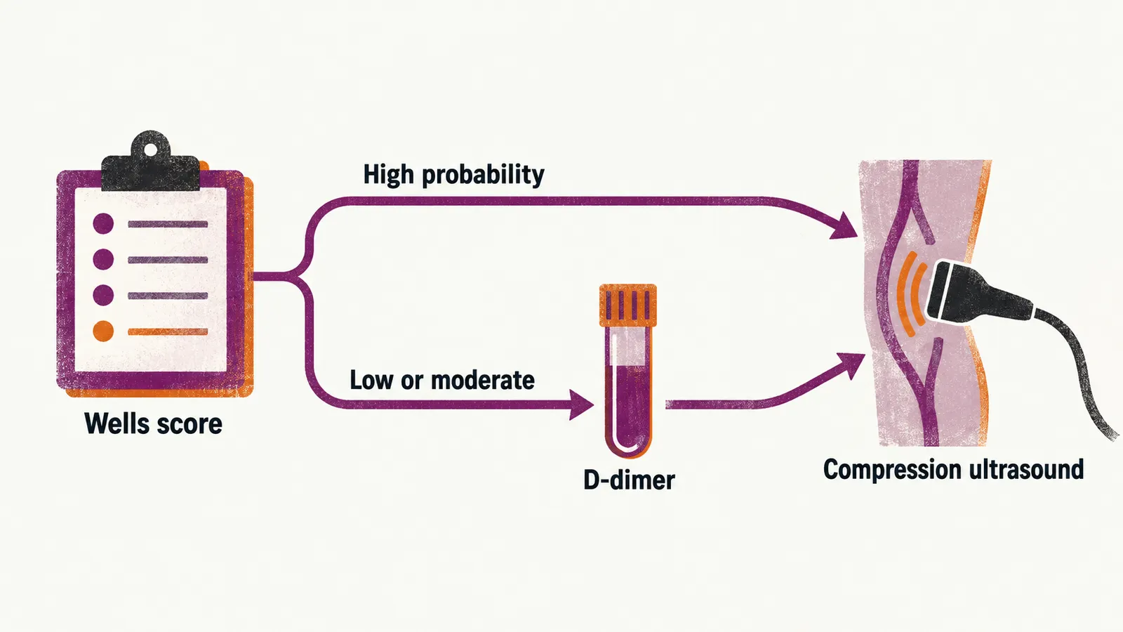
For PE the same principle applies. The Wells score for PE sets the clinical probability; when PE is unlikely and the D-dimer is negative, PE is ruled out, whereas when PE is likely, or the D-dimer is positive, imaging is needed to confirm it. Computed tomographic pulmonary angiography (CTPA) is the imaging test of choice, with ventilation–perfusion scanning used when contrast cannot be given.
Once DVT or PE is confirmed, two questions follow: which anticoagulant to give, and whether the event reflects an inherited tendency to clot that is worth testing for.