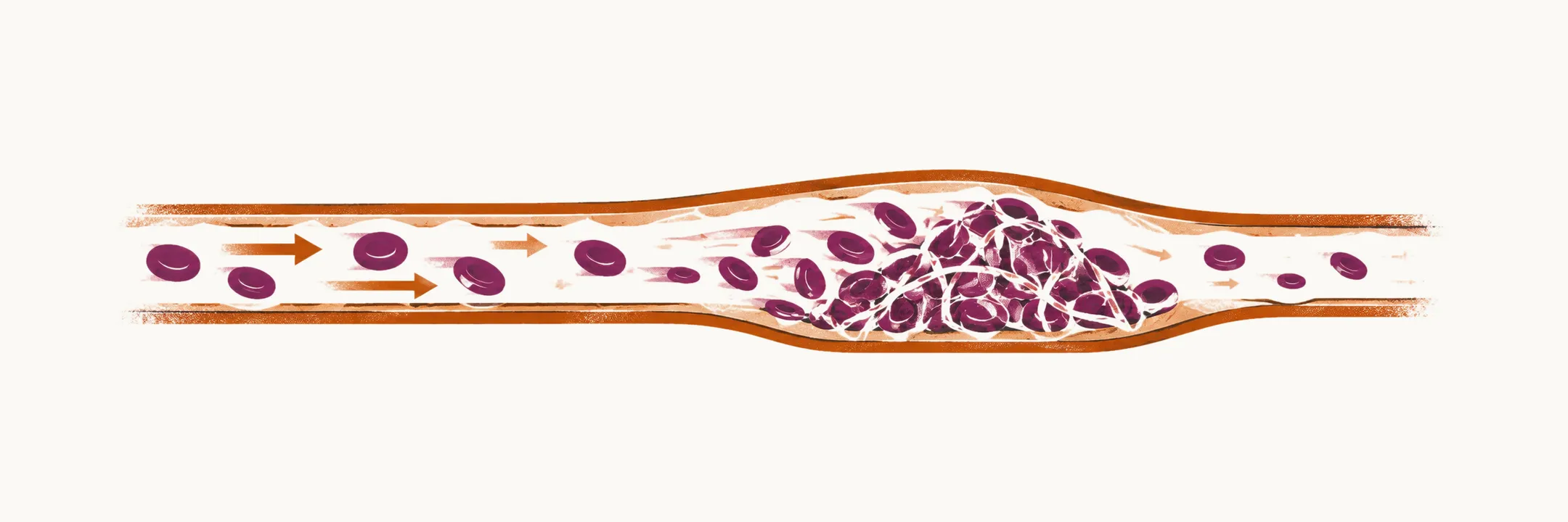
Venous thrombosis is the formation of a thrombus in a vein. Its primary form is deep-vein thrombosis or DVT, and the subsequent embolisation of that thrombus in the lung is pulmonary embolism or PE. Together they are called venous thromboembolic disorder, or VTE.
More than 90% of VTE occur as lower-extremity DVT and PE, but venous thrombosis is possible in other venous parts of the body.
Venous blood flows slowly and at low pressure, so a venous thrombus is rich in fibrin and red cells and forms with less dependence on platelets than an arterial one. That is why venous thrombosis is driven mainly by a hypercoagulable state (an increased tendency of the blood to clot, from abnormal coagulation or fibrinolysis), by stasis of blood flow, or by both.
Some of the main risk factors for venous thrombosis are:
- a hypercoagulable state
- immobilisation and venous stasis
- orthopedic, abdominal and neurological surgery, all significant risk factors
- community- or hospital-acquired infection
- hormone replacement therapy (HRT) and oral contraceptives
- pregnancy
Genetics of venous thrombosis
Genetic causes are a less common reason for VTE, but they are the usual reason a patient is tested. They are easiest to follow against the body’s natural anticoagulants: antithrombin inhibits thrombin and factor Xa, and activated protein C, with its cofactor protein S, cleaves and inactivates factors Va and VIIIa. Inherited defects fall into two groups: loss-of-function mutations that weaken these natural anticoagulants, and gain-of-function mutations that increase a procoagulant.
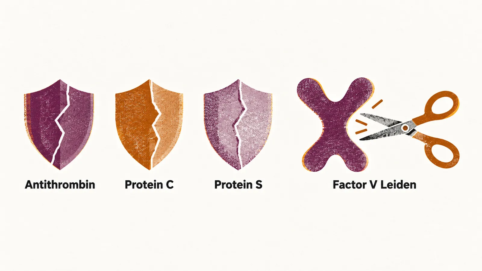
Deficiencies of the natural anticoagulants
Antithrombin deficiency is a rare autosomal dominant condition (one altered copy of the gene is enough to cause it) in which the main inhibitor of thrombin and factor Xa is reduced. It appears in two forms: type I, with a low antigen level (too little antithrombin protein), and type II, with a normal antigen level but low activity (the protein is present but works poorly). Antithrombin deficiency can also be acquired, as in nephrotic syndrome, liver disease and disseminated intravascular coagulation (DIC).
Protein C deficiency is autosomal dominant and reduces the cleavage of factor Va and factor VIIIa. The rare homozygous or compound heterozygous forms, in which both copies of the gene are affected, can lead to fatal purpura fulminans, while the heterozygous forms give a moderate risk of thrombosis. It can also be acquired.
Protein C deficiency is also a setting for warfarin-induced skin necrosis. In the first days of warfarin, the fall in protein C and factor VII outpaces that of the other vitamin K-dependent factors, producing a short-lived hypercoagulable state.
Protein S deficiency is likewise autosomal dominant. Protein S is the cofactor that allows activated protein C to inactivate factor Va and VIIIa, so its loss raises clotting risk in the same way.
Gain-of-function variants
Factor V Leiden is the most common inherited thrombophilia. It removes the site at which activated protein C cleaves factor Va, so factor Va resists inactivation — a state called activated protein C resistance — and clotting is favoured. It is inherited in an autosomal dominant pattern; heterozygous carriers (one copy) are common, and homozygous carriers (two copies) carry a higher risk. The diagnosis uses a functional test based on a mixture of the patient’s plasma and standard plasma without factor V Leiden, and a PCR test that detects the variant in DNA.
The G20210A variant of the prothrombin gene is another gain-of-function change: it leads to an elevated level of factor II (prothrombin), so more thrombin can be generated.
Fibrinolysis and thrombosis
Clotting risk depends not only on how readily clots form but also on how readily they are broken down. Fibrinolysis is the process that breaks a clot down, and it is driven by tissue plasminogen activator (t-PA), which converts plasminogen to plasmin, the enzyme that digests fibrin. Plasminogen activator inhibitor (PAI-1) inhibits t-PA, so a high PAI-1 level reduces fibrinolytic capacity and is associated with an increased risk of thrombosis. In these states t-PA is often elevated as well, because it rises with the endothelial activation that accompanies the same conditions, but the net effect is reduced clot breakdown.