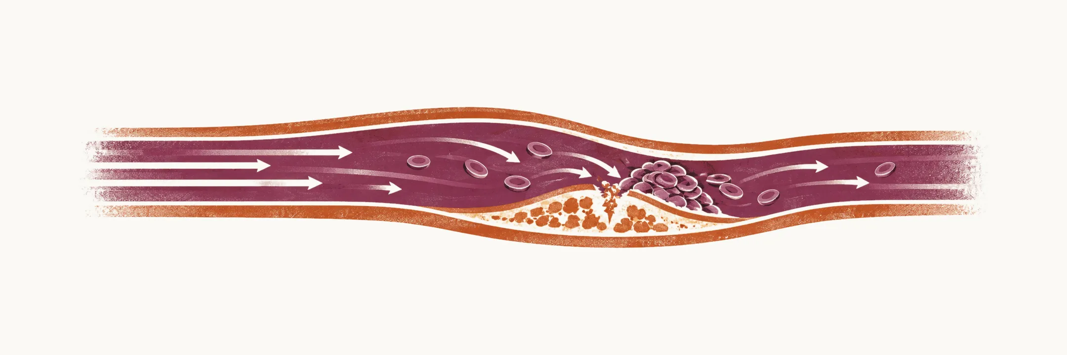
In arterial thrombosis, the main factors related to the formation of thrombus are platelet abnormalities and vessel wall abnormalities. In essentially all forms of arterial thrombosis, platelets adhere to the vessel wall and recruit other platelets, which leads to the formation of thrombus in arterial regions.
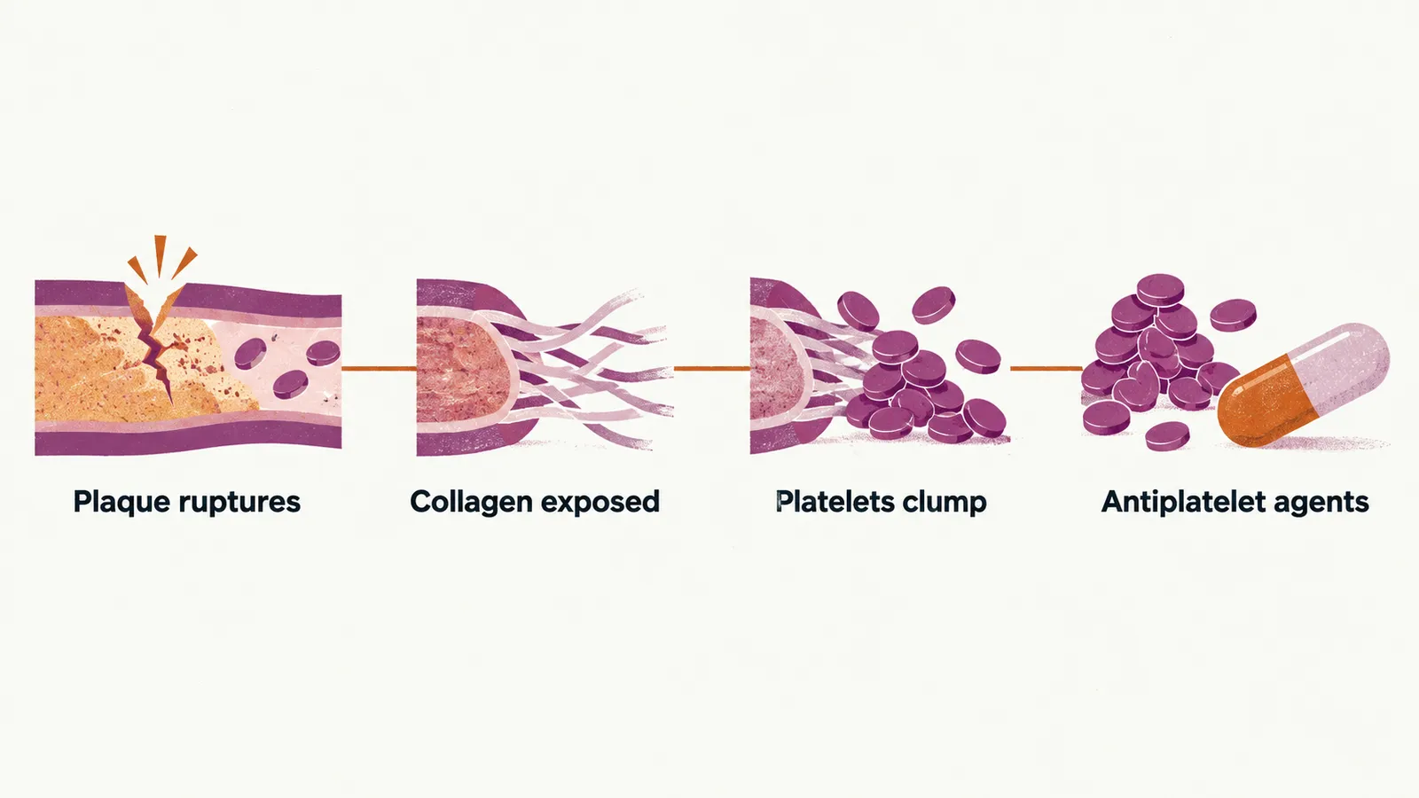
Arteries carry blood at high velocity under high shear, the frictional force that fast-flowing blood exerts on the vessel wall, and the thrombus that forms in them is built differently from a venous one. Injury to the vessel wall exposes collagen and von Willebrand factor, a protein that anchors platelets to the damaged wall, from beneath the endothelium, the cell layer that lines the vessel. The fast flow favors platelet adhesion and aggregation over the formation of fibrin, the protein mesh of a clot. Most arterial thrombi begin where an atherosclerotic plaque, a fatty deposit in the arterial wall, has ruptured.
Because platelets are the dominant cell in an arterial thrombus, the drugs used to prevent arterial events are antiplatelet agents such as aspirin and the P2Y12 inhibitors, which block one of the platelet’s receptors for ADP, rather than the anticoagulants that treat venous thrombosis.
Risk factors
Since most arterial thrombi start at a ruptured plaque, conditions that injure the arterial wall, raise platelet reactivity, or promote clotting raise the risk. There are several different risk factors for arterial thrombosis:
- smoking
- a high level of low-density lipoprotein (LDL) cholesterol
- diabetes
- pregnancy
- chemotherapy agents
- systemic lupus erythematosus (SLE)
- rheumatoid arthritis (RA)
Antiphospholipid syndrome
SLE and RA bring autoimmune disease into that list, and SLE is also one of the settings of antiphospholipid syndrome (APS). APS is an acquired autoimmune thrombophilia, an increased tendency to thrombosis caused by the patient’s own antibodies, and it is related to both arterial and venous thrombosis. APS can be primary, occurring on its own, or secondary to SLE or other autoimmune disorders.
APS can be seen in three forms:
- thrombotic APS, in which arterial, venous or small-vessel thrombosis occurs
- obstetric APS, defined by one or more unexplained fetal deaths at or after 10 weeks of gestation, a premature birth before 34 weeks because of preeclampsia, eclampsia or placental insufficiency, or three or more consecutive miscarriages before 10 weeks
- catastrophic APS, in which small-vessel thrombosis affects three or more organs within one week and carries a high mortality
The antibodies in APS are directed against phospholipid-binding proteins rather than against phospholipid itself. They reduce the activation of protein C, a natural anticoagulant, and its ability to inactivate factors Va and VIIIa, and they promote the binding of prothrombin (factor II) to phospholipid surfaces, so the balance shifts toward clotting.
The diagnosis of APS depends on detecting these antibodies, and some laboratory tests can be used:
- anticardiolipin antibody (ACL) by ELISA (enzyme-linked immunosorbent assay)
- anti-β2 glycoprotein I antibodies
- lupus anticoagulant assay (PTT LA)
The lupus anticoagulant shows up as a prolonged activated partial thromboplastin time (aPTT), a clotting-time test. The prolongation is not corrected by mixing with normal plasma, but it is corrected by adding phospholipids.
The antibodies must persist to establish the diagnosis: they are detected on two occasions at least 12 weeks apart.