
Amyloid angiopathy presents in older adults with bleeding in cortical territories, the opposite distribution to hypertensive small-vessel disease. When an older patient shows lobar microbleeds or a peripheral haemorrhage without a clear hypertensive mechanism, amyloid angiopathy belongs at the top of the differential.
Haemorrhagic manifestations
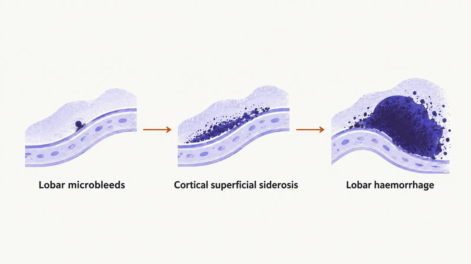
Bleeding from the fragile vessels ranges from tiny leaks to a large haematoma, and the smallest signs come first. Lobar microbleeds are the earliest sign. They are tiny bleeds, invisible on CT and visible only on blood-sensitive MRI sequences, often multiple and all in lobar positions.
Repeated small surface bleeds leave a trace. Cortical superficial siderosis is hemosiderin, the iron-containing residue of old blood, deposited along the brain surface from prior small surface haemorrhages, and it is visible on susceptibility-weighted imaging (a blood-sensitive MRI sequence).
Lobar intracerebral haemorrhage is the most dramatic presentation: bleeding in peripheral regions such as the frontal, parietal, or occipital lobes rather than the basal ganglia or brainstem.
Transient focal neurological episodes are a further haemorrhage-related syndrome: brief, recurrent, stereotyped spells that can resemble transient ischaemic attacks. They occur in a subset of patients, often alongside cortical superficial siderosis, and are a recognised presenting syndrome in the current diagnostic criteria.
Non-haemorrhagic manifestations
Not every feature comes from bleeding. White matter hyperintensities are bright areas on FLAIR MRI (fluid-attenuated inversion recovery) that reflect chronic ischaemia and vascular injury, overlapping in appearance with hypertensive small-vessel change. Enlarged perivascular spaces, particularly in the centrum semiovale (the deep white matter of the cerebral hemispheres), reflect impaired perivascular amyloid drainage.
Progressive cognitive decline can dominate the picture, often mixed with Alzheimer pathology given how frequently the two conditions coexist.
An older patient with a lobar haemorrhage, multiple lobar microbleeds, no deep haemorrhagic lesions, and cognitive or white matter change fits amyloid angiopathy until another cause is established. Confirming that impression depends on what blood-sensitive MRI shows and on how the findings are graded.