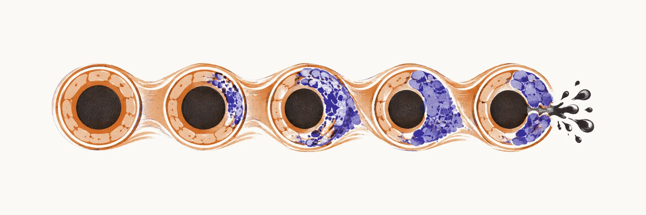
Amyloid-beta is a protein that deposits in the walls of small and medium-sized cortical and leptomeningeal arteries (the arteries of the cortex and of the membranes covering the brain surface). It accumulates along the perivascular drainage pathways, the channels alongside vessels by which the cortex normally clears amyloid, which is why the disease affects outer-brain vessels and spares the deep perforating arteries supplying the basal ganglia.
Why the vessel wall fails
Amyloid deposition replaces the smooth muscle layer of the vessel wall. The wall loses both structural support and the ability to autoregulate, that is, to adjust vessel calibre and keep flow steady when blood pressure changes, so it becomes stiff and fragile. Small leaks appear first as microbleeds, tiny bleeds detectable only on blood-sensitive MRI sequences. Larger lobar haemorrhages follow when a weakened wall gives way completely. Severely affected vessels can develop fibrinoid necrosis, a destructive wall change associated with a higher risk of rupture.
The shared biology with Alzheimer disease
Alzheimer disease is the better-known amyloid disease, and the two conditions involve the same protein deposited in different compartments: Alzheimer disease deposits amyloid-beta as parenchymal plaques (within brain tissue), while amyloid angiopathy deposits it in vessel walls. Overlap is common, and many patients carry elements of both diseases. The ε4 allele of the APOE gene (apolipoprotein E) raises the risk of each, consistent with impaired amyloid clearance from the brain as a shared mechanism.
Vessel-wall failure on one side and coexisting Alzheimer pathology on the other shape what patients actually show, both bleeding and cognitive decline.