
Cerebral amyloid angiopathy (CAA) means amyloid-beta deposited in the walls of small brain vessels. CAA-related inflammation is the treatable face of this vessel disease. It is a rare subacute encephalopathy, a diffuse disturbance of brain function, not an acute bleed. Headache, seizures, behavioural or cognitive change, and focal signs evolve over weeks. MRI shows asymmetric subcortical T2 oedema (swelling seen as bright signal on T2-weighted images) overlying cortex loaded with amyloid. Most patients also carry lobar microbleeds. First-episode frequencies run roughly 58% cognitive or behavioural change, 42% headache, 42% seizure, and 19% focal deficit.
The tempo is the bedside clue. Bleeding strikes suddenly with a focal deficit. Inflammation smoulders for weeks with encephalopathy.
Diagnosis without biopsy
Probable disease can be diagnosed from clinical and MRI features alone. The clinicoradiological criteria reach about 82% sensitivity (the share of true cases identified) and 97% specificity (the share of non-cases correctly excluded) for probable disease (about 82% and 68% for possible disease). Biopsy remains the gold standard but is increasingly replaced in practice. CSF (cerebrospinal fluid) anti-amyloid-beta autoantibody assays are research tools, not validated diagnostics.
Treatment and response
High-dose corticosteroids are first-line treatment, often with a taper. Cyclophosphamide or mycophenolate is added in selected cases. Treated patients improve far more often than untreated ones: clinical improvement in about 94% versus 50%, radiographic improvement in about 86% versus 29%. Recurrence is also less likely with immunosuppression (26% versus 71%). These comparisons come from retrospective cohorts, and half of untreated patients improve spontaneously, so the figures describe association rather than a tested protocol. No standardised regimen exists.
ABRA, the vasculitic pole
Amyloid-beta-related angiitis (ABRA) is the vasculitic pole of the same spectrum. In CAA-related inflammation the infiltrate sits around vessels without destroying them. In ABRA, inflammation invades the vessel wall, often with granulomas. The two are separable only by biopsy. ABRA affects younger patients, shows leptomeningeal enhancement, and behaves like primary CNS vasculitis. It responds to the same immunosuppression. Whether the two are one spectrum or separate entities is unresolved.
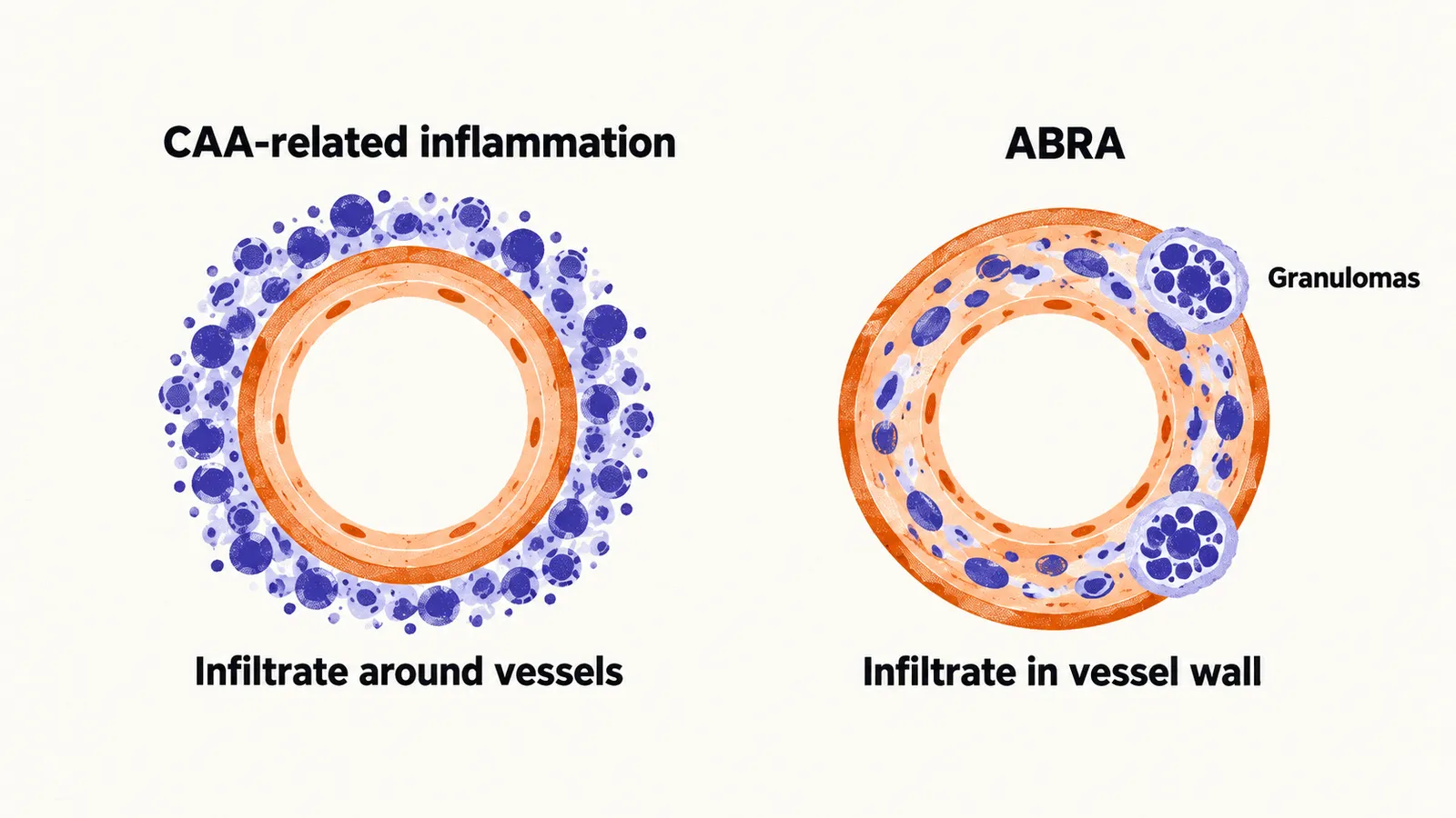
The practical point is directional. Ordinary amyloid angiopathy management avoids anything that raises bleeding risk. Inflammatory disease needs the opposite: immunosuppression. Confusing the two withholds the one effective treatment.
For the usual bleeding form, the remaining question is what a first haemorrhage predicts over the following years.