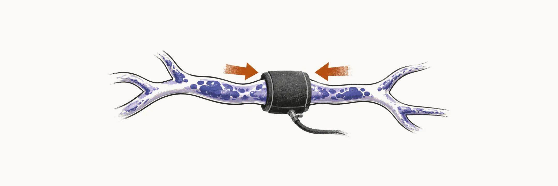
There is no disease-modifying therapy for amyloid angiopathy. Management is entirely about preventing the next haemorrhage, and every decision is shaped by how fragile the affected vessels are. That covers avoiding antithrombotic drugs (antiplatelet agents and anticoagulants), limiting mechanical stress on the vessels through blood pressure control, handling an existing haematoma, and avoiding anti-amyloid antibodies in patients whose vessels are already affected.
Avoiding antithrombotics
Antiplatelet agents and anticoagulants raise the bleeding risk from amyloid-laden vessels, and an anticoagulated microbleed can become a catastrophic lobar haemorrhage. They are therefore generally avoided once probable disease is established. The difficult case is coexistence with atrial fibrillation or vascular disease, where a thrombotic risk also demands attention. That decision is individualised, weighing stroke risk against haemorrhage risk, with the microbleed burden on blood-sensitive MRI informing the bleeding side of the scale.
Blood pressure control
Tight blood pressure management reduces the mechanical stress on fragile vessels and is the main modifiable measure against rebleeding, following standard post-haemorrhage secondary-prevention guidance. Acute hypertensive episodes can precipitate rupture, so both long-term control and avoidance of sharp rises matter.
Surgical considerations
Once a bleed has happened, the question is whether to remove the haematoma. Evacuation of a lobar haematoma follows standard intracerebral haemorrhage decision-making by location, size, and consciousness level. The surrounding tissue in amyloid angiopathy is itself diseased, which raises the risk of perioperative rebleeding. Tissue biopsy is rarely performed but can establish pathology-supported diagnostic certainty where management truly depends on it.
Anti-amyloid antibodies and ARIA
Anti-amyloid antibodies for Alzheimer disease, such as lecanemab, mobilise amyloid that is also present in vessel walls. They carry a risk of amyloid-related imaging abnormalities (ARIA): oedema or effusion type (ARIA-E) and haemorrhage type including microbleeds and superficial siderosis (ARIA-H).
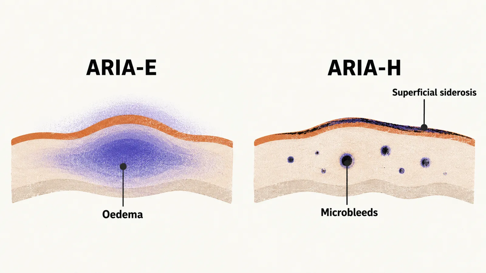
Patients with amyloid angiopathy or a substantial baseline haemorrhagic lesion burden face higher ARIA risk, so MRI screening for such lesions is required before starting treatment, and extensive CAA-type findings weigh against it. The likelihood that a given patient carries substantial vessel amyloid, by age and genotype, therefore becomes a practical question.