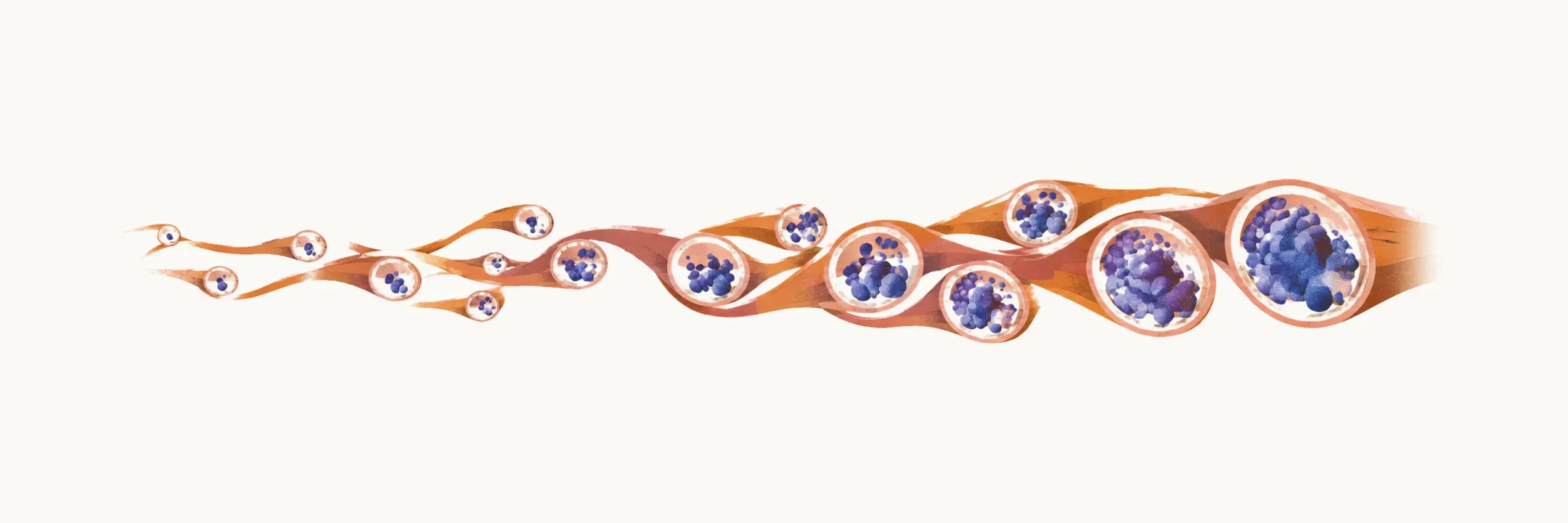
Cerebral amyloid angiopathy, amyloid-beta deposited in the walls of small brain vessels, is a disease of ageing. Sporadic disease, meaning the usual non-inherited form, usually appears from the fifth decade onward. Moderate-to-severe vessel pathology is found in about one quarter of the general elderly population at autopsy. That figure comes from cohorts with a mean age near 85, so it does not describe prevalence at age 60.
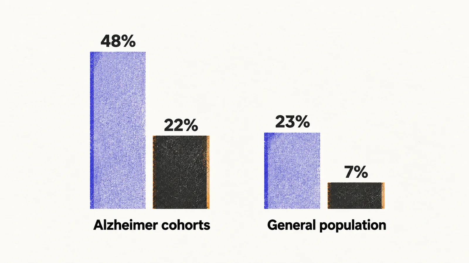
MRI sees only part of the disease. Lobar microbleeds are small bleeds confined to the cortical territories, and they are the MRI marker of vessel pathology. In Alzheimer cohorts, pathology prevalence is roughly double the rate of strictly lobar microbleeds on MRI (48% versus 22%). In the general population the gap is roughly threefold (23% versus 7%). Microbleeds on MRI likely identify only severe vessel disease.
Pre-test probability, the likelihood of the disease before testing, follows the bleed location. About half of lobar haemorrhages are driven by amyloid angiopathy, by pathology (57%) and by Boston-criteria diagnosis (about 50%). The Boston criteria are the clinical and MRI-based framework used to diagnose the disease. In unselected intracerebral haemorrhage the share is roughly one fifth to one quarter. In cognitively normal older adults, moderate-to-severe vessel pathology affects about 6%.
Genotype adds a second layer of risk. The gene for apolipoprotein E (APOE) has an epsilon-4 and an epsilon-2 allele, and they separate two steps of the disease. The epsilon-4 allele increases amyloid deposition in vessel walls and brings the first lobar haemorrhage forward by roughly 5 years. The epsilon-2 allele instead raises the risk of bleeding from vessels already loaded with amyloid, through destructive wall changes such as fibrinoid necrosis. Epsilon-2 is not protective overall.
Hereditary forms
Hereditary forms are rare and autosomal dominant, meaning one mutated copy of the gene is enough to cause disease, and they are usually caused by APP gene mutations (the gene for the amyloid precursor protein). They begin earlier and run a more severe course than sporadic disease. The Dutch type (APP E693Q) is the best studied. Carriers develop recurrent strokes in the fifth and sixth decades. Stroke is often the first sign and is fatal in about one third. Survivors commonly develop dementia, and about half of those with strokes develop recurrent seizures. Far rarer non-APP forms include Icelandic (cystatin C) and British/Danish (ITM2B) disease.
Both sporadic and hereditary disease centre on bleeding and cognitive decline. A rare inflammatory variant instead evolves over weeks.