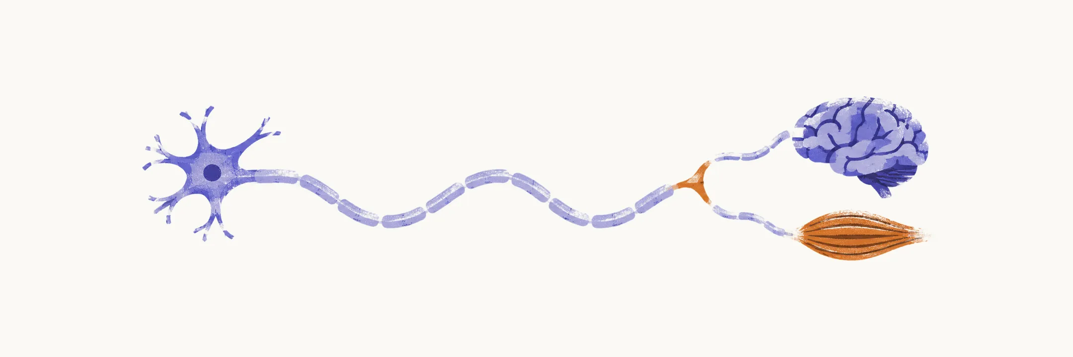
A floppy baby is an infant with abnormally low muscle tone (hypotonia): reduced resistance to passive stretch, so the baby feels limp when held, rests in splayed positions such as the frog-leg posture, and shows signs such as head lag, slipping through on vertical suspension, and draping on ventral suspension. Hypotonia is a sign, not a disease. The task is to localize where along the motor pathway, from cortex to muscle, the signal is being lost.
The distinction that organizes everything is central versus peripheral. Central means the brain and spinal cord pathways above the anterior horn cell, the spinal motor neuron whose axon runs to muscle. Peripheral means the lower motor unit: the anterior horn cell itself, the peripheral nerve, the neuromuscular junction, and the muscle. In central hypotonia the infant looks floppy but still generates movement when stimulated, and reflexes are normal or brisk. In peripheral hypotonia the infant is floppy and weak in proportion, with diminished or absent reflexes. Central causes are more common overall, but several peripheral causes have disease-modifying treatments, so mislocalizing a peripheral condition as a central one can cost a therapeutic window.
Choose a route through the topic
The route begins with the method. Floppy Baby Examination covers the bedside assessment that makes the central-versus-peripheral call: observation before touching, the general examination for dysmorphic and systemic clues, tone maneuvers such as the traction response and suspension, and what reflex patterns mean. Floppy Baby Differential Diagnosis then turns that call into a list of causes: central and peripheral causes, conditions that mimic hypotonia, and the diagnoses that must not be missed.
Three causes show the distinction at work at different levels of the motor pathway. Spinal Muscular Atrophy is the most important treatable peripheral cause: the gene loss behind it, the backup gene that modifies severity, the clinical picture, and the three disease-modifying therapies. Congenital Myotonic Dystrophy is a mixed cause, in which muscle and brain are both affected: why myotonia is absent at birth, and why the mother should be examined and tested. Hypotonic Cerebral Palsy is the main central cause: why an early brain injury presents as floppiness first and spasticity later, and how to separate it from motor unit disease.
Floppy Baby Diagnostic Workup explains which test to order once the examination has localized the lesion, from blood genetics and creatine kinase (CK) rules to imaging, neurophysiology, and biopsy. The last two causes are ones where that testing logic does much of the work. Pompe Disease covers the floppy infant with an enlarged heart: the cardiac and ECG clues, blood enzyme and genetic confirmation, and why treatment timing and CRIM status, an immunologic test done before treatment, matter. Prader-Willi Syndrome covers neonatal hypotonia with poor feeding that later reverses into excessive eating, and why diagnosis starts with a methylation test.
Start with the examination if the bedside assessment is unfamiliar, and read onward in the order above: each step builds on the localization made before it.