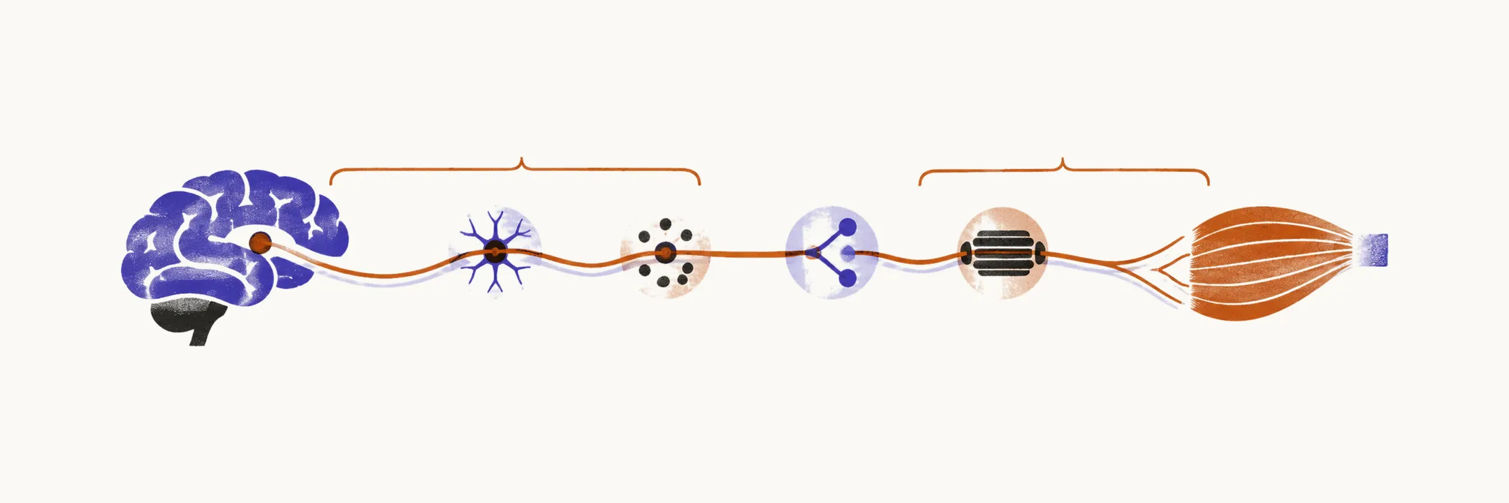
Every differential for the floppy infant starts from anatomy: at which level of the motor pathway, from cortex to muscle, is the problem? Central causes lie in the brain or spinal cord above the anterior horn cell, the spinal motor neuron whose axon runs to muscle. Peripheral causes lie in the lower motor unit: the anterior horn cell, peripheral nerve, neuromuscular junction, or muscle. Central causes are more common, accounting for roughly 60–80% of cases compared with 15–30% peripheral, but the peripheral group contains the treatable diagnoses, so the split must be made deliberately rather than by probability alone.
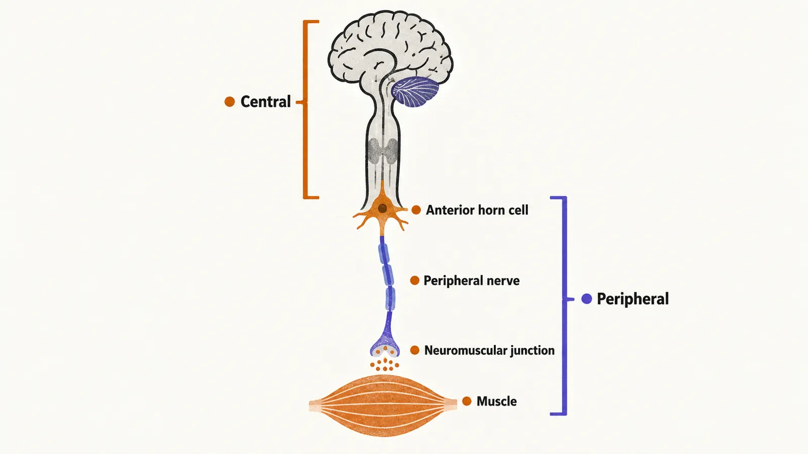
The central versus peripheral comparison
Central hypotonia is also called non-paralytic, because the infant is floppy but relatively strong; peripheral hypotonia is called paralytic, because the floppiness comes with true weakness.
| Feature | Central (non-paralytic) | Peripheral (paralytic) |
|---|---|---|
| Movement when stimulated | Relatively preserved; floppy but reactive | Poor; floppy and weak in proportion |
| Deep tendon reflexes | Normal, brisk, or persistent primitive reflexes | Diminished or absent |
| Alertness and cognition | Often reduced; poor tracking, lethargy | Often alert despite severe weakness |
| Associated clues | Microcephaly, seizures, dysmorphic features, organ malformations | Fasciculations, atrophy, weak cry, respiratory failure |
For example: an infant with microcephaly, absent visual tracking, and brisk reflexes localizes centrally, toward a structural brain problem imaged with MRI, because anterior horn cell or muscle disease does not shrink the head or quicken reflexes. Conversely, tongue fasciculations (fine twitching of muscle fibres visible under the surface) with absent reflexes and a bell-shaped chest from intercostal weakness localize to the anterior horn cells.
Central causes
Hypoxic-ischemic encephalopathy (HIE), brain injury from reduced oxygen and blood supply around birth, is the most common single central cause. It is usually obvious from the perinatal history of difficult delivery or neonatal depression. The diagnoses that must be actively considered are therefore the ones without a clear perinatal event:
- chromosomal conditions, of which Down syndrome is the most common chromosomal cause
- Prader-Willi syndrome: hypotonia with characteristic facial features, feeding difficulty, reduced reflexes, and hypogonadism
- intracranial hemorrhage
- structural brain malformations
- inborn errors of metabolism
Hypotonic cerebral palsy is the main chronic central cause: perinatal injury to the corticospinal tracts, the pathways that carry movement commands from the cortex to the spinal cord, produces flaccidity first and spasticity over months.
Some central pictures are temporary. Sepsis, maternal drug exposure, congenital hypothyroidism, and prematurity (judged against corrected gestational age, counted from the expected due date) can each produce a centrally floppy picture that improves when the underlying condition is treated.
Peripheral causes
The three peripheral causes to recognize first are spinal muscular atrophy, congenital myotonic dystrophy, and Pompe disease. Pompe disease has a clue of its own: cardiac enlargement on chest radiograph in a hypotonic neonate is a useful pointer toward it.
Beyond these three, the differential covers the rest of the lower motor unit: peripheral neuropathies at the nerve, neuromuscular junction disorders (transient neonatal myasthenia, infantile botulism) where nerve signals pass to muscle, and congenital myopathies and congenital muscular dystrophies in the muscle itself. An acute ascending flaccid weakness after an infection in an older infant points instead to Guillain-Barre syndrome, a different time course from the congenital floppy presentation.
Systemic conditions that mimic hypotonia
The temporary central pictures above shade into a wider group. A systemically unwell infant can look floppy without any primary motor unit disease; listed causes are congenital heart disease, sepsis, hypothyroidism, rickets, malabsorption, malnutrition, and renal tubular acidosis. In these infants weakness is marginal, some movement persists on stimulation, and tone recovers when the systemic problem is corrected. Connective tissue disorders such as Ehlers-Danlos syndrome produce joint hypermobility through collagen rather than through reduced neural tone, and need to be distinguished on that basis.
Diagnoses that must not be missed
Five conditions are traditionally singled out because each carries specific management or counseling consequences: Down syndrome, Prader-Willi syndrome, Pompe disease (look for cardiac involvement), Zellweger spectrum disorder, and spinal muscular atrophy. What unites them is not anatomy but actionability: each changes what the clinician does next, from cardiac surveillance to family counseling to disease-modifying therapy.