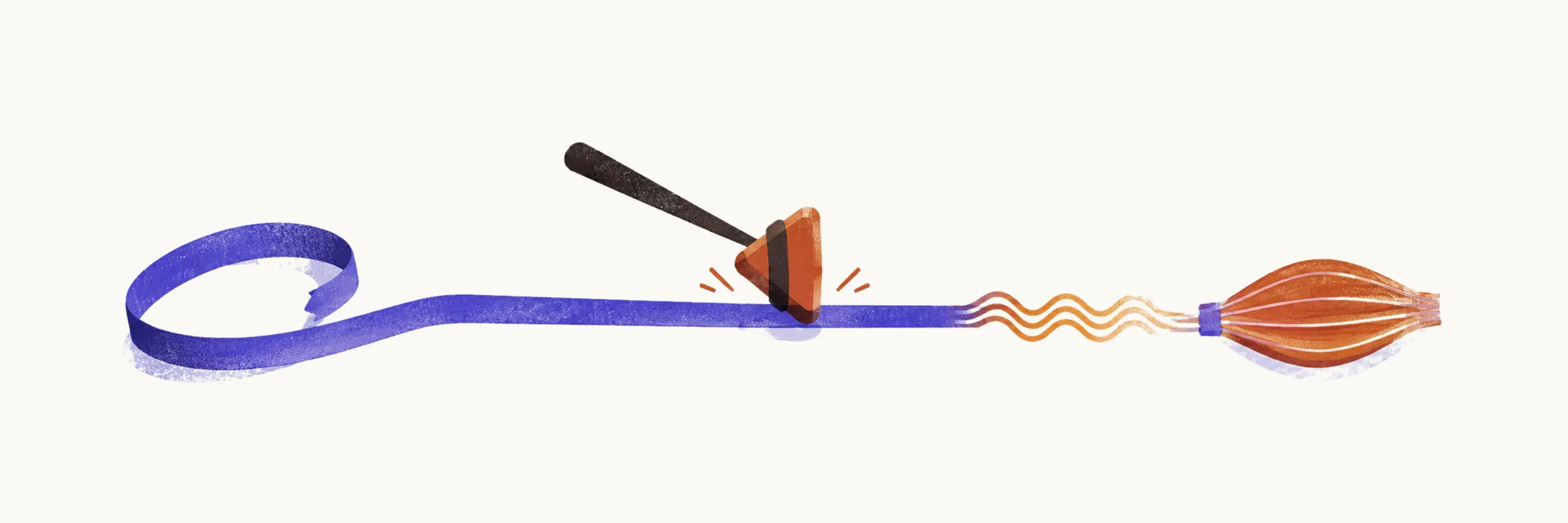
The physical examination is the decisive tool in a floppy infant: it confirms that tone is truly reduced and gives the first answer to the central-versus-peripheral question. Central means the problem lies in the brain or spinal cord above the anterior horn cell, the spinal motor neuron whose axon runs to muscle. Peripheral means it lies in the lower motor unit: the anterior horn cell, peripheral nerve, neuromuscular junction, or muscle.
Tone means resistance of muscle to stretch, while weakness means reduced strength. The examination tests each separately, because their relationship is what localizes the lesion.
Observation before touching
Watch first. A healthy neonate is restless, with shifting limb movements and a reactive face. A hypotonic infant lies still with limbs splayed, typically in full abduction and external rotation of the legs with flaccid extension of the arms (the frog-leg posture). Note asymmetry between sides, which suggests a focal lesion.
Note alertness as well: peripherally weak infants often look alert despite profound weakness, while centrally affected infants may seem lethargic with poor visual tracking. Lack of spontaneous movement already suggests weakness, and the cry, facial expression, sucking, and respiratory effort each reflect strength in different muscle groups.
General examination
Observation shows how the infant moves; the general examination looks for what else is wrong. Examine the undressed infant, because dysmorphic features (unusual physical features that suggest a syndrome) and systemic clues point toward central or syndromic causes.
Work from the head down. At the face, look at facial proportions, palate shape, and whether the mouth stays closed at rest; jaw weakness that leaves the mouth hanging open is a visible clue in congenital myotonic dystrophy. At the chest, check symmetry and shape, since a thin, poorly expanding chest reflects weakness of the intercostal muscles between the ribs. Then listen to the heart, because cardiac enlargement can hide behind respiratory signs and points toward conditions such as Pompe disease.
Below the chest, organ enlargement, genital anomalies, dislocated hips, and fixed joint contractures (arthrogryposis) each carry localizing meaning. Contractures are the paradoxical clue in a floppy infant: they signal severe hypotonia that began before birth, because a fetus that never moves develops stiff joints.
Tone maneuvers
Once the infant has been watched and examined, handling tests tone directly. Each maneuver tests postural tone, the antigravity resistance normal muscle provides. The first three move the whole infant against gravity and show whether the head and trunk hold; the last two move a single limb across a joint and feel for resistance.
- Traction response (pull to sit). Pull the infant symmetrically by the wrists toward sitting. Some head lag is normal in the first weeks, but a healthy infant visibly works to bring the head up; a floppy infant shows no resistance and the head hangs back.
- Vertical suspension. Lift under the axillae without gripping the chest. A normal baby feels solid, while a floppy baby slips through the hands for lack of axial tone.
- Horizontal (ventral) suspension. Rest the infant face-down along the forearm. A normal baby tenses trunk and limbs, while a floppy baby drapes over the arm with head and limbs hanging.
- Scarf sign. Draw one arm across the chest around the opposite shoulder. Absent resistance, with the elbow crossing the midline effortlessly, indicates hypotonia.
- Heel to ear. Flex hip and knee to bring the foot toward the ear. A floppy infant folds with almost no resistance, except where arthrogryposis has fixed the joints.
These maneuvers show whether tone is reduced. They do not show where the problem lies; the reflexes begin to answer that.
Reflexes
Deep tendon reflexes are harder to elicit in neonates, which makes their pattern more informative than any single result. Brisk reflexes or clonus (rhythmic, repeated contractions after a sudden stretch) point above the anterior horn cell, toward a central cause. Absent or very depressed reflexes point to the lower motor unit, as in spinal muscular atrophy. Normal reflexes occur in mild central hypotonia and in systemic illness where the reflex arc is intact.
Primitive reflexes, automatic movement patterns present in the newborn that the maturing brain normally suppresses, add a second axis. The asymmetric tonic neck reflex (brief arm extension on the face side when the head turns) is transient in healthy infants. When it persists rigidly for as long as the head is held turned, cortical inhibition of primitive patterns is impaired, which favors a central cause.
Putting the examination together
Two questions close the bedside assessment. First, is hypotonia truly present: draping, slipping through, a positive scarf sign, and excessive joint mobility leave no ambiguity. Second, does movement match the floppiness: preserved antigravity movement with brisk reflexes leans central, while proportional weakness with absent reflexes leans peripheral.