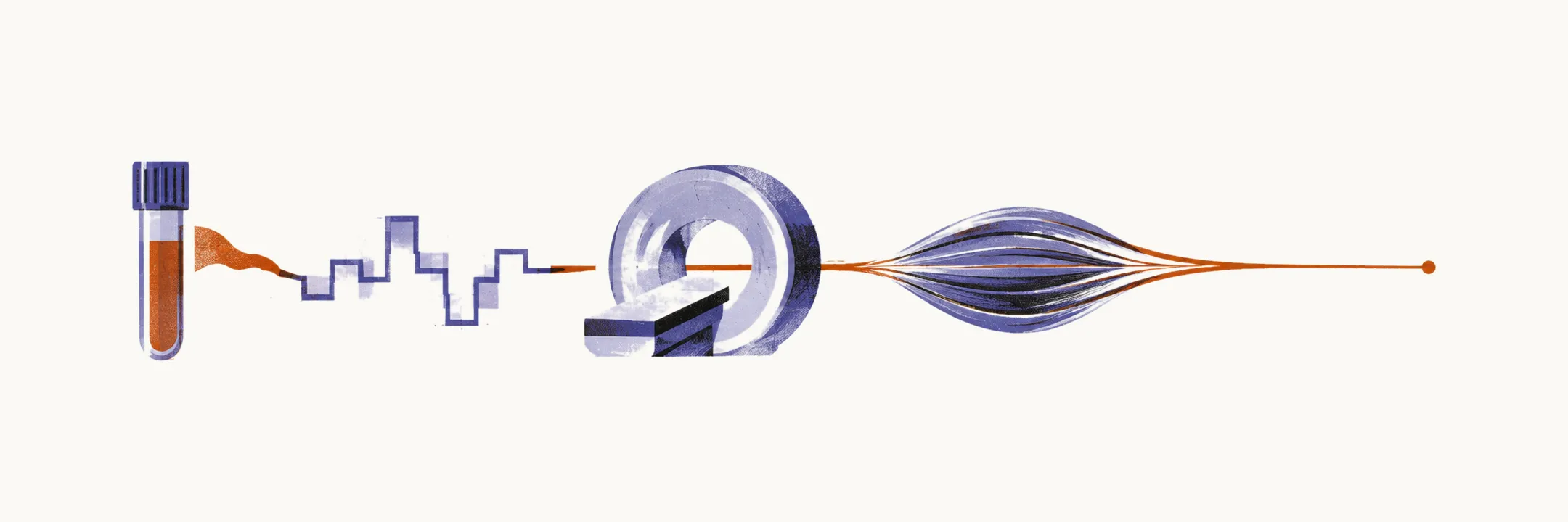
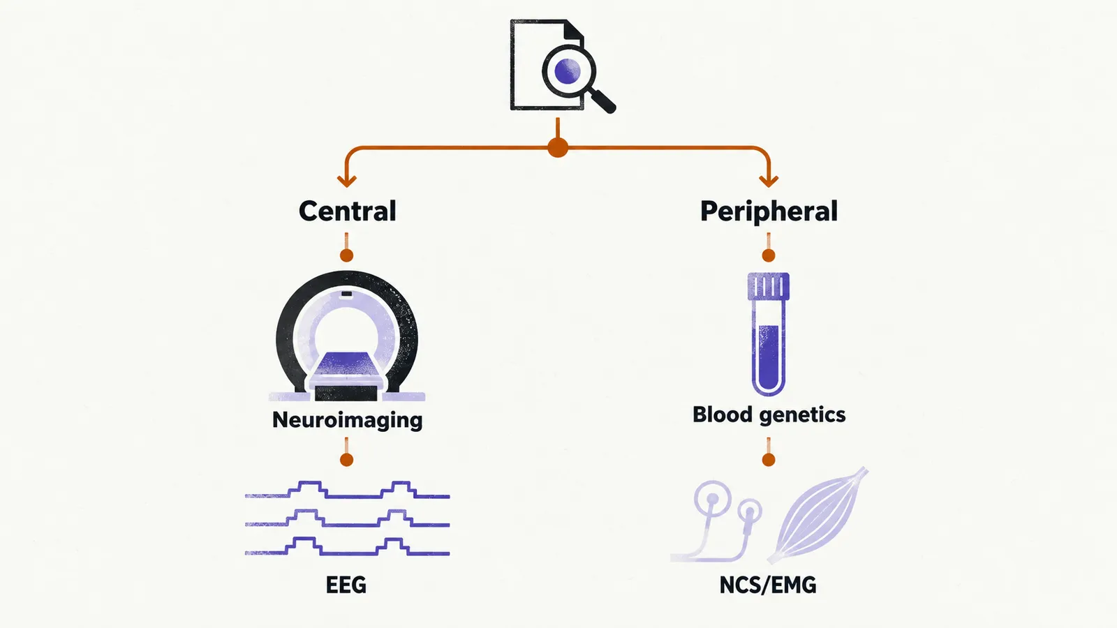
Testing follows localization. The major task in evaluating a hypotonic neonate is determining whether the pathology sits above the anterior horn cell (central) or in the lower motor unit (peripheral), because each branch orders different tests. The anterior horn cell is the spinal motor neuron whose axon runs to muscle; the lower motor unit is that neuron together with its peripheral nerve, the neuromuscular junction, and the muscle. At the bedside, a centrally floppy infant still moves when stimulated and has normal or brisk reflexes, while a peripherally floppy infant is weak in proportion to the floppiness, with diminished or absent reflexes.
The central branch runs through neuroimaging, EEG (electroencephalography, a recording of the brain’s electrical activity), and metabolic or genetic testing. The peripheral branch runs through creatine kinase (CK), an enzyme that leaks into the blood from damaged muscle; targeted blood genetics; nerve conduction studies with electromyography (NCS/EMG), which record electrical activity in nerve and muscle; and muscle biopsy.
Exclude acute central and systemic causes first
In neonatal series, central causes account for roughly two-thirds of cases, with hypoxic-ischemic encephalopathy (HIE), brain injury from reduced oxygen and blood supply around birth, the most common single cause. This proportion comes from regional data and predates broad genomic testing, so treat it as orientation rather than a constant. The practical consequence stands: excluding acute central and systemic causes such as HIE, sepsis, and hypoglycaemia is the first move.
First-line blood tests when the picture is peripheral
When the examination points peripherally, targeted blood tests come before any invasive study. First-line peripheral investigations are:
- CK
- DNA testing for myotonic dystrophy
- SMN1 gene-deletion testing, for spinal muscular atrophy
- an EDTA blood sample (the anticoagulated tube used for DNA) for Prader-Willi testing
- chest radiograph with echocardiography, to look for cardiomegaly or cardiomyopathy
- microarray CGH (comparative genomic hybridization), which detects missing or extra chromosomal material
Interpreting CK in the newborn
CK needs cautious interpretation in the newborn. Levels tend to be higher at birth, rise across the first 24 hours, and increase with acidosis. A high initial value therefore proves nothing on its own and should be repeated. No numeric cutoff defines myopathy (primary muscle disease) here.
Keep NCS, EMG, and biopsy second-line
Blood genetics can end the search before any invasive test. SMN1 deletion testing is high-yield because the deletion is present in 95% of cases of spinal muscular atrophy type 1. This figure is scoped to type 1 and does not generalize to all spinal muscular atrophy. A positive SMN1 result settles the diagnosis, and invasive neurophysiology then adds nothing.
When blood tests leave the question open, NCS/EMG can separate neurogenic, myopathic, myotonic, and myasthenic patterns, but the study is difficult in the first 6 months of life. Muscle biopsy goes to histology, immunohistochemistry, electron microscopy, and respiratory-chain analysis as a second-line step. Two stopping rules govern both: positive blood genetics obviate biopsy, and a normal early EMG does not exclude disease.