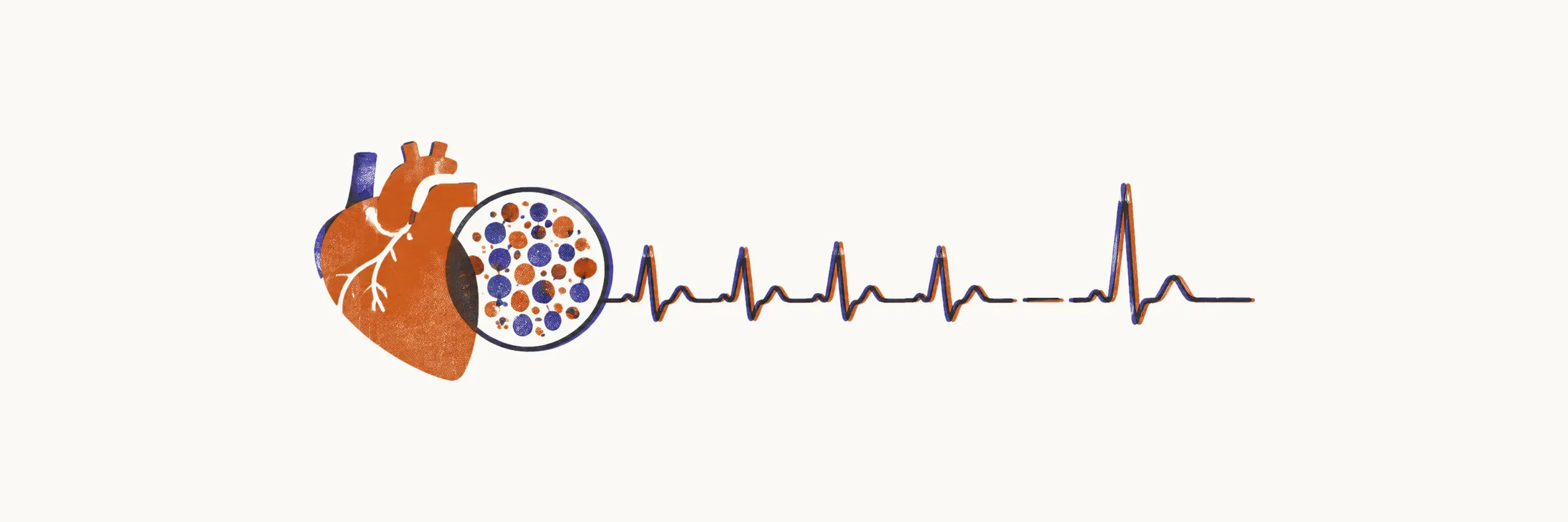
Pompe disease is an autosomal recessive deficiency of the lysosomal enzyme acid alpha-glucosidase (GAA), which breaks down glycogen inside lysosomes, the cell’s digestive compartments. Without it, glycogen accumulates most prominently in skeletal, cardiac, and smooth muscle. Because skeletal and heart muscle are both affected, the classic infant is floppy and has a diseased heart, and that pairing is the clue that points toward the diagnosis.
The infantile cardiac clue
Classic infantile disease presents with prominent cardiomegaly, weakness, and hypotonia. Cardiomegaly and hypertrophic cardiomyopathy (thickened heart muscle) are usually identified in the first weeks of life and progress toward left ventricular outflow obstruction, in which the thickened wall narrows the path blood takes out of the left ventricle, with diminished lung volume. When untreated, the disease is fatal in infancy, with death commonly occurring in the first two years from cardiopulmonary insufficiency.
The electrocardiogram adds a clue: a short PR interval with very tall QRS complexes. The short PR interval appeared in 14 of 19 infantile cases in one small historical series, so it supports suspicion without ruling anything out. The working chain is a floppy infant plus cardiomegaly or hypertrophy plus a short-PR ECG pointing at Pompe disease.
CK is supportive, never diagnostic
Creatine kinase (CK), an enzyme released from damaged muscle, is often measured early in a floppy infant. CK elevation is a sensitive but very nonspecific marker of Pompe disease. The greatest elevation is found in infantile-onset patients, historically as high as 2000 UI/L. That number reflects the assay and era behind it, so it serves as context rather than a cutoff. CK supports the suspicion and never makes the diagnosis.
Confirm with blood enzyme testing plus genetics
The diagnosis is established by GAA enzyme deficiency in isolated lymphocytes or mixed leukocytes, by biallelic pathogenic GAA variants (disease-causing changes in both copies of the gene) on molecular testing, or both. Current practice thus rests on blood-based enzyme activity, molecular testing, or both, reserving fibroblast assay for selected cases.
Dried-blood-spot GAA testing reaches close to 100% sensitivity for infantile disease, and combined with urinary Glc4, a glycogen breakdown product excreted in urine, it can effectively rule the disease out. Fibroblast culture was the diagnostic gold standard in 2006 but takes weeks to return, so follow the current hierarchy: blood enzyme plus GAA genetics first, with no invasive biopsy required for diagnosis.
Treat early, and check CRIM status first
The speed of that confirmation matters because treatment works best early. Enzyme replacement therapy (ERT), regular intravenous infusions of the missing enzyme, should start as soon as infantile-onset or symptomatic disease is diagnosed. Infants treated before 6 months of age and before needing ventilation show improved survival, ventilator-independent survival, reduced cardiac mass, and better motor-skill acquisition than untreated infants. These outcome data are observational, compared against historical controls, so the idea that days matter stands as a principle rather than a measured dose-response.
CRIM (cross-reactive immunologic material) status, meaning whether the infant makes any GAA protein at all, even a nonfunctional one, should be determined before the first infusion. CRIM-negative infants generally develop high-titer antibodies against the replacement enzyme during ERT and need immunomodulation built into the plan early, optimally before the first infusion, which calls for a specialist centre.
Testing of all at-risk siblings of any age is warranted so that early diagnosis allows early treatment.
Pompe disease shows how one extra sign beyond the floppiness, here the heart, can steer the diagnosis. In other floppy infants the extra sign is feeding: poor suck in the newborn, then a changed relationship with food in early childhood.