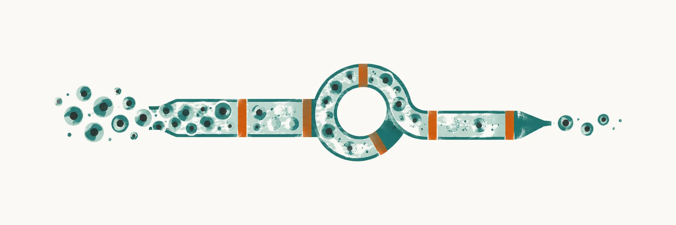
Childhood acute lymphoblastic leukemia (ALL) is the most common childhood cancer, a disease in which immature lymphoid precursors, called blasts, displace normal blood-cell production. It is treated with combination chemotherapy delivered in protocol phases, and the intensity of the whole program is set by the risk group assigned at diagnosis and by the early response to treatment.
Where the modern approach started
The first useful drug in childhood ALL came from a failed hypothesis. In the 1940s Sidney Farber reasoned that folic acid, known to drive marrow recovery in malnourished patients, would help children with leukemia. The opposite happened: folate appeared to accelerate the leukemic process. That observation became the question — if folate feeds the disease, what happens if the supply is cut off? — and the antifolate aminopterin produced the first temporary remissions in childhood acute leukemia. Antifolate therapy, and then single-agent chemotherapy in general, became the platform for everything that followed.
From that starting point, drugs accumulated one at a time: mercaptopurine and methotrexate in the 1940s and 1950s, corticosteroids, vincristine and L-asparaginase in the 1950s and 1960s, cytarabine and cyclophosphamide in the 1960s, anthracyclines in the 1960s, epipodophyllotoxins in the 1970s, and later imatinib, clofarabine, nelarabine and blinatumomab. The lasting lesson is not the date of any one drug. It is that each new agent was inserted into a protocol backbone that already existed — and it is the backbone, not the individual drug, that cures ALL.
The treatment backbone
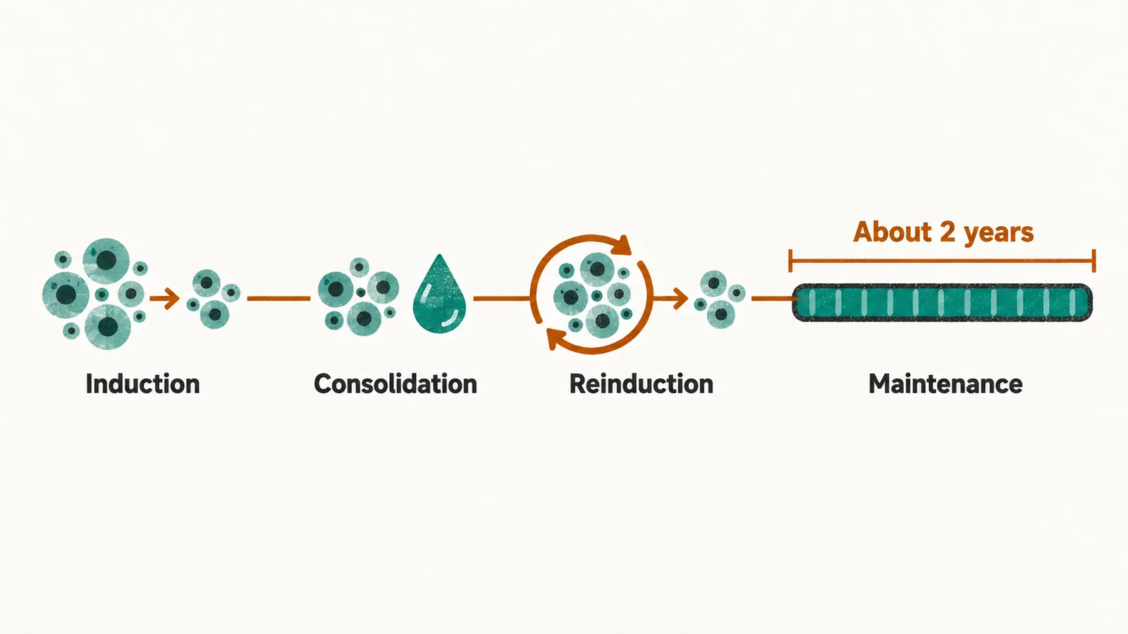
The backbone developed by the Berlin-Frankfurt-Münster (BFM) group is built from four phases, with the middle blocks repeated as protocol cycles.
- Induction (protocol IA). Vincristine, a corticosteroid (prednisone or dexamethasone), L-asparaginase, and an anthracycline such as daunorubicin. The goal is remission — a marrow with fewer than 5% blasts and recovering peripheral counts, conventionally assessed around day 33.
- Consolidation (protocol IB). Cyclophosphamide, cytarabine, mercaptopurine, and intrathecal methotrexate, meaning methotrexate injected into the cerebrospinal fluid. The goal is to deepen the remission while treating the central nervous system, which chemotherapy given systemically reaches poorly.
- Reinduction or delayed intensification (protocol II). A repeat of the induction strategy with several agents added. The goal is to attack any residual disease again, with a different drug combination.
- Maintenance (protocol M). Daily oral mercaptopurine with weekly methotrexate, plus periodic vincristine and corticosteroid pulses, continued for about 2 years from the start of therapy. The goal is to suppress regrowth of any dormant clone.
Intrathecal prophylaxis and high-dose methotrexate
Because systemic chemotherapy reaches the central nervous system poorly, central nervous system prophylaxis is delivered intrathecally, at lumbar puncture, on a fixed schedule. In BFM-based protocols the methotrexate dose is age-banded — around 8 mg for children aged 1 to under 2 years, 10 mg for 2 to under 3 years, and 12 mg from 3 years onward — because cerebrospinal fluid volume tracks age more closely than body surface area does.
High-dose methotrexate is given at 2 or 5 g/m² per course (doses per square metre of body surface area), followed by leucovorin (folinic acid) rescue at set doses and hours after the infusion starts. Leucovorin supplies the reduced folate that methotrexate blocks, and giving it after the infusion rather than with it is what makes the high dose tolerable: normal tissues such as marrow and mucosa recover while much of the antitumor effect has already been established. In the AIEOP-BFM schedule for the 5 g/m² dose, leucovorin 15 mg/m² is given at hours 42, 48 and 54 after the start of the infusion, or 7.5 mg/m² when the levorotatory form (levoleucovorin) is used. During the high-dose blocks, oral mercaptopurine continues at about 25 mg/m²/day.
Measurable residual disease
The backbone is the same for every child; how hard it is pushed is not, and the measurement that has come to decide it is measurable residual disease (MRD): the leukemic cells still present after treatment has started, at a depth microscopy cannot reach. It is the in vivo test of whether treatment is working. Immunoglobulin and T-cell receptor gene rearrangements are unique to each leukemic clone, so they can serve as targets for the polymerase chain reaction (PCR), a method that amplifies a chosen DNA sequence until it can be detected. PCR can find one leukemic cell among 10,000 or more normal cells — a sensitivity of better than 10⁻⁴. Flow cytometry, which reads the markers carried by individual cells, is the method used for the earliest timepoint.
The prognostic weight is the reason it now drives protocols. Work published in the late 1990s established that residual disease detectable after induction was a stronger predictor of relapse than age, white cell count or cytogenetics at presentation, and later trials confirmed the relationship at a threshold of 10⁻³ (one leukemic cell in a thousand) after consolidation. Timepoints are typically day 15 by flow cytometry, day 33 (end of induction), and around week 12 (before or at the end of consolidation). Children with no detectable disease do well; children with persistent disease are intensified and considered for transplantation.
Risk stratification
Not every child needs the same intensity, and modern protocols assign risk from three kinds of information together: baseline cytogenetics (the chromosomal and genetic lesions of the leukemic clone at diagnosis), early response, and measurable residual disease.
The AIEOP-BFM ALL 2000 scheme, from the joint Italian (AIEOP) and BFM trial group, is a convenient reference point:
- Standard risk meant no high-risk features, with a negative MRD result at both timepoints.
- Medium risk meant no high-risk features but positive residual disease after induction.
- High risk meant any of: prednisone poor response (blasts persisting in the blood after the initial prednisone phase), BCR::ABL1-positive disease (the Philadelphia chromosome) or KMT2A-rearranged disease, failure to reach remission by day 33, or residual disease at or above 10⁻³ after consolidation.
Within the high-risk group, sub-strata determined which children were offered allogeneic transplantation — stem cell transplantation from a donor — in first remission.
The 2009 revision simplified the algorithm around the same variables — lineage, high-risk cytogenetics (hypodiploidy, a leukemic clone with too few chromosomes, was added to the high-risk criteria in this revision), and residual disease measured by flow cytometry at day 15. It also made a further change with long-term consequences: cranial irradiation was replaced in most children by intensified systemic cyclophosphamide and intensified intrathecal methotrexate, with prophylactic cranial irradiation reduced to selected subgroups and to a lower dose (12 Gy, in place of the historical 18–24 Gy; the gray, Gy, is the unit of absorbed radiation dose) where it is still used.
Five shifts have accumulated since 2000 and explain most of the improvement in outcome:
- residual disease, rather than age and white cell count alone, decides intensity;
- cranial irradiation replaced by intensified systemic and intrathecal therapy;
- dedicated protocols for infants and for BCR::ABL1-positive disease (with a tyrosine kinase inhibitor added to chemotherapy);
- intensified dexamethasone and pegylated asparaginase, given as PEG-asparaginase 2500 IU/m² every 2 weeks in place of native E. coli asparaginase;
- residual disease–based indications for stem cell transplantation in first remission.
The AIEOP-BFM ALL 2000 trial enrolled about 4,800 children aged 1–17 years and reported 5-year outcomes of roughly 80% event-free survival (survival without relapse or another adverse event) and 90% overall survival, an improvement on the pre-2000 figures for the same disease.
The 2017 generation
The successor protocol, AIEOP-BFM ALL 2017, was built on a revised residual-disease stratification that treats early and final results differently according to biological subgroup. It also asks a randomized question in high-risk disease: whether two cycles of post-consolidation immunotherapy with blinatumomab, an antibody construct that links the patient’s T cells to CD19 on leukemic B cells, given at 15 µg/m²/day by continuous infusion for 28 days, can replace two conventional high-intensity chemotherapy courses. The rationale is the trade-off that runs through the whole field — the need for new treatment elements that reduce treatment-related mortality and morbidity while holding the cure rate.
What the cure leaves behind
Cure is not the end of the clinical story. The drugs and the intensified schedules that produce these survival figures leave behind health problems that appear years later — osteonecrosis, anthracycline cardiomyopathy, secondary malignancy, thrombosis — and the survivors who carry them will live with the consequences for decades.