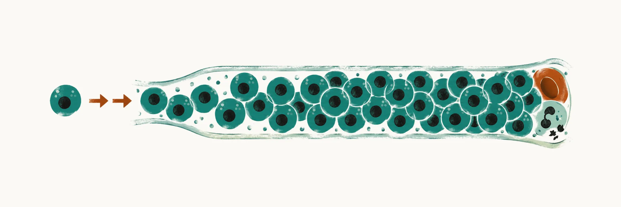
Leukemia is the malignant transformation and clonal proliferation of hematopoietic precursor cells — the immature marrow cells that normally give rise to blood cells. Clonal means that the leukemic cells all descend from a single transformed cell. The clone fails to mature, so the cells that accumulate are blasts — immature precursors arrested at an early stage. They fill the bone marrow, displace normal hematopoiesis (blood-cell production), and spill into the peripheral blood, where a blood film can show them.
Why leukemia heads the list in childhood
Cancer in childhood is rare — around 1% of all cancers — but in high-income countries it is the leading cause of death from disease in children, and leukemia is its most common form, accounting for roughly 30% of childhood cancers, close to one in three. Within childhood acute leukemia, about 80% of cases are acute lymphoblastic leukemia (ALL) and 15–20% are acute myeloid leukemia (AML), and chronic leukemias are uncommon enough that the word leukemia in a child nearly always means an acute one.
The contrast with adults runs through the whole topic. In adults, myeloid leukemias are relatively more prominent and chronic leukemias are common; in children the lymphoid, acute end of the spectrum dominates. A high lymphocyte count in a child is also far more often reactive than leukemic, and the blood film together with the rest of the count usually separates the two. When a chronic myeloid picture does appear in a child, BCR::ABL1-positive chronic myeloid leukemia is the entity to think of.
Some children carry a higher risk from birth. Children with Down syndrome have a markedly increased risk of acute leukemia, with acute megakaryoblastic leukemia (a leukemia of the precursors of platelet-producing cells) over-represented, a form of childhood acute myeloid leukemia with a biology and treatment of its own.
Two axes of classification
Leukemia is classified two ways at once, and both axes carry information.
| Axis | Options | What it decides |
|---|---|---|
| Tempo | Acute — blasts accumulate quickly, illness over days to weeks. Chronic — slower expansion, illness over months. | Almost all childhood leukemia is acute. |
| Lineage | Lymphoblastic (lymphoid precursor) or myeloid (myeloid precursor). | The ALL-versus-AML distinction, which sets the treatment backbone and the markers used to confirm the diagnosis. |
Lymphoblastic and lymphocytic name the same lineage; the acute precursor neoplasms are called lymphoblastic in the World Health Organization (WHO) classification. ALL is the most common childhood type, which is why most of the treatment literature in this field is a literature about ALL.
The clinical picture comes from the marrow
Almost all of the presenting signs are consequences of a marrow that is full of blasts. Normal blood-cell production is crowded out, and the same clone infiltrates tissue.
- Anemia — pallor, fatigue, lassitude, breathlessness on exertion.
- Neutropenia — fever and infection, sometimes with systemic signs that look disproportionate to the apparent source.
- Thrombocytopenia — bruising, petechiae, mucosal bleeding, prolonged bleeding from minor cuts.
- Tissue infiltration — hepatosplenomegaly is the most common physical finding, with lymphadenopathy, testicular enlargement in boys, and bone or joint pain that can present as a limp or refusal to walk.
The spleen and lymph nodes are not primarily diseased. They are involved by the same clone that occupies the marrow, and the marrow is where the diagnosis is made.
Infiltration can also be dangerous in its own right. In T-cell ALL the mediastinal mass is the feature to keep in mind: it can compress the trachea or the superior vena cava, so a child with a wide mediastinum, breathlessness and facial or upper-limb swelling is a clinical urgency rather than an outpatient referral.
Diagnosis: count, film, then marrow
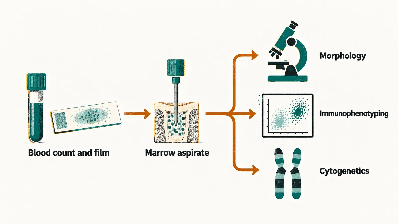
A peripheral blood count and film come first. They may be entirely suggestive — anemia, neutropenia, thrombocytopenia, and circulating blasts — but a normal film does not exclude leukemia, and the diagnosis rests on the bone marrow aspirate. One aspirate supplies three independent lines of information.
- Morphology. Light microscopy describes what the blasts look like. The historical French-American-British (FAB) scheme divided ALL into L1, L2 and L3; modern classification is biology-based and integrates the two lines below rather than relying on appearance alone.
- Immunophenotyping by flow cytometry. Flow cytometry reads the marker proteins carried by individual cells. Surface and cytoplasmic markers, named by CD (cluster of differentiation) numbers, assign the blasts to B-cell lineage (CD19, CD22, CD79a, CD10) or T-cell lineage (CD2, CD3, CD7), and identify the maturation stage at which the clone is arrested. This is what distinguishes B-ALL from T-ALL from AML on a single sample.
- Cytogenetics and molecular genetics. Recurrent genetic abnormalities define biological subtypes, carry prognostic weight, and increasingly determine treatment.
A blast count of 20% or more in the marrow or blood is the conventional threshold for acute leukemia. WHO classification then carves out exceptions: certain recurrent lesions — PML::RARA, RUNX1::RUNX1T1, CBFB::MYH11, and some KMT2A rearrangements among them — are definitional, so the diagnosis can be made at a lower blast count when one of them is present. This matters because such children are treated as acute leukemia even though the marrow looks less involved.
Cytogenetics: the findings that change prognosis
The third line of information from the aspirate, the genetics of the blasts, is also where a few findings change prognosis. Two conventions make them readable. A translocation such as t(12;21) is an exchange of material between two chromosomes, here 12 and 21. When the exchange joins two genes, the resulting fusion gene is written with a double colon, as in ETV6::RUNX1. Two translocations recur in every discussion of prognosis:
- t(12;21) — ETV6::RUNX1 (formerly TEL-AML1). The most frequent chromosomal alteration in childhood B-cell precursor ALL, found in roughly 20–25% of cases. It is cryptic on karyotype — invisible on standard chromosome analysis — and needs FISH (fluorescence in situ hybridization) or molecular testing to detect, and it carries a favorable prognosis.
- t(9;22) — BCR::ABL1, the Philadelphia chromosome. Historically the worst-prognosis pediatric ALL subtype; it is now redirected to therapy with tyrosine kinase inhibitors, with substantially better outcomes than before those drugs existed.
Chromosome number matters as well as chromosome structure. Hyperdiploidy (more than 50 chromosomes) is favorable, while hypodiploidy (too few chromosomes) is adverse, as are KMT2A (MLL) rearrangement and iAMP21 (intrachromosomal amplification of chromosome 21).
Prognosis: why children with ALL do comparatively well
Reported outcomes in children treated on contemporary protocols are of the order of 80% event-free survival (alive without relapse or another defined event) and above 90% overall survival, with some series reporting overall survival in the mid-90s. The reason is part biological and part therapeutic.
- Biology. Favorable subtypes such as ETV6::RUNX1, hyperdiploidy and TCF3::PBX1 are common in children and rare in adults.
- Response-adapted therapy. Treatment intensity is assigned by baseline biology and by measurable residual disease — leukemic cells still detectable by sensitive laboratory methods after microscopy can no longer see them — early in therapy, so the favorable group receives less treatment and less toxicity while high-risk children are intensified.
What turns that favorable biology into these outcomes is the treatment built around it: the same features decide how much of that treatment each child needs, together with how quickly the disease responds to the first phase of it.