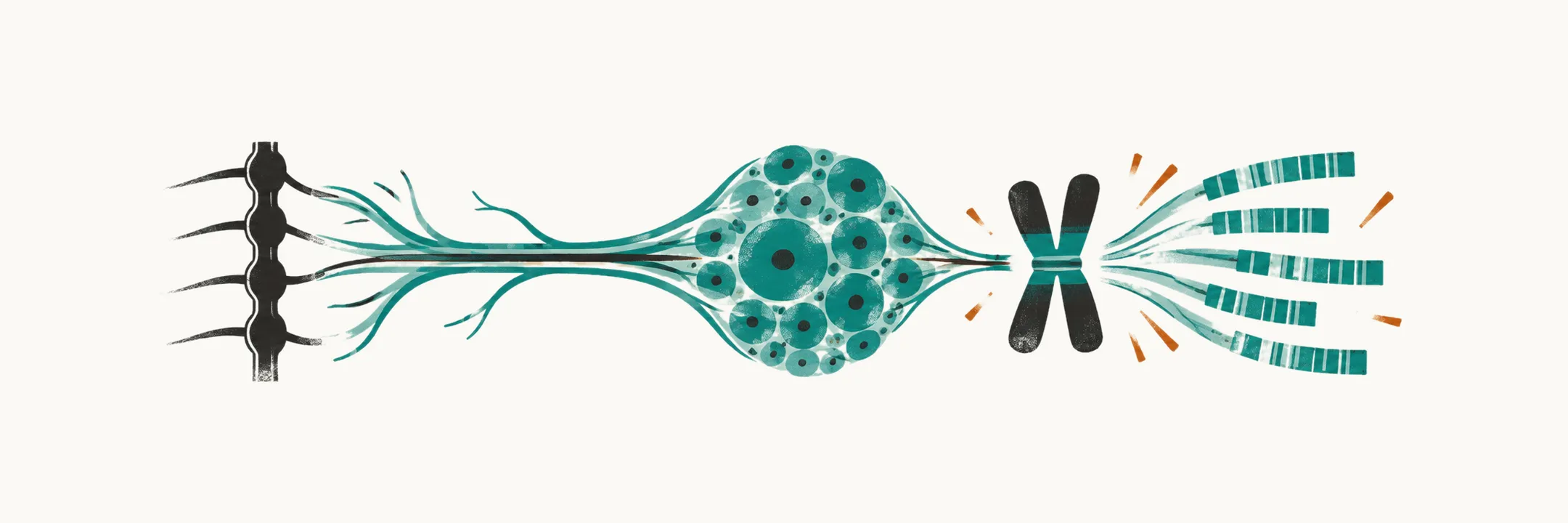
Neuroblastoma is a malignant embryonal tumor derived from precursor cells of the sympathetic nervous system — the adrenal medulla and the sympathetic ganglia. It is the most common solid tumor outside the brain in early childhood and the most common cancer diagnosed in infancy, and it behaves in ways no leukemia does: it can regress without treatment in a baby, and it kills older children despite the most intensive therapy in pediatrics.
One cell of origin, a family of tumors
Because the tumor arises from embryonal sympathetic tissue, it sits in a family organized by how much the cells have differentiated:
- Ganglioneuroma — fully differentiated, benign.
- Ganglioneuroblastoma — intermediate, mixed differentiation.
- Neuroblastoma — undifferentiated and malignant.
- Pheochromocytoma — a tumor of the catecholamine-secreting chromaffin cells of the adrenal medulla, which shares the sympathetic origin but is far more common in adults than in children.
The naming follows the same convention as nephroblastoma (Wilms tumor), medulloblastoma and retinoblastoma: a malignant tumor of tissue that should have finished differentiating before birth.
The infant paradox
Neuroblastoma behaves in two opposite ways depending on the age of the child. In infants under 1 year it can regress spontaneously, or differentiate into a benign ganglioneuroma, without treatment. In older children it is one of the most aggressive solid tumors in pediatrics. The same disease, at the same anatomical site, can be self-limited in a baby and lethal in a school-age child.
The biology behind spontaneous regression is not fully established. Differences in neurotrophin receptor signaling are part of the explanation — favorable infant tumors tend to express high levels of the nerve growth factor receptor TrkA, while aggressive tumors signal through other neurotrophin pathways — and immune mechanisms and telomere maintenance have also been proposed. What is settled is the clinical consequence: age at diagnosis is one of the strongest prognostic factors in the disease.
Epidemiology
Neuroblastoma accounts for roughly 7–8% of all childhood cancers and is the most frequent malignancy of infancy. Approximately 90% of cases are diagnosed before the age of 5 years, and more than a third are diagnosed during the first year of life; the median age at diagnosis falls in the second year. That age distribution is why an abdominal mass in a young infant should raise the possibility immediately.
Clinical presentation
An abdominal mass is only one way in. What the child shows depends on where the primary tumor sits, how far it has spread, and whether it secretes anything, so the signs fall into three groups: local effects, systemic effects, and paraneoplastic syndromes.
Local effects, by primary site. Most tumors arise in the abdomen (the adrenal medulla or sympathetic ganglia being the commonest single site), followed by the posterior mediastinum, pelvis and neck.
- An abdominal tumor causes distension, a palpable mass, or pressure symptoms. Within the abdomen the main alternative to keep in mind is Wilms tumor, which is renal rather than adrenal or paravertebral and usually presents in the preschool years; imaging usually separates the two, and so does the tumor-marker pattern.
- A posterior mediastinal or paravertebral tumor can compress the trachea, causing cough and dyspnea, or grow through an intervertebral foramen into the spinal canal as a dumbbell tumor that compresses the cord.
- Tumors in the neck or apex of the chest can compress the sympathetic chain and produce Horner syndrome — ptosis, miosis and enophthalmos.
- Periorbital involvement gives the yellow-brown ecchymoses known as raccoon eyes, which can be mistaken for injury.
Systemic effects of marrow involvement and tumor burden. Metastatic disease is present at diagnosis in a substantial proportion of children — around 60% overall — and is more common in children over 1 year, in whom the usual sites are bone marrow, bone, liver and skin. Marrow involvement and tumor burden produce weight loss, fever, pallor, lassitude, irritability, bone and joint pain, and cytopenias. Bone pain can present simply as a child who will not stand or walk — a sign worth taking seriously in a toddler. Infants may present with bluish subcutaneous nodules, the so-called blueberry muffin lesions of metastatic skin involvement.
Paraneoplastic syndromes are effects produced at a distance from the tumor, by antibodies or secreted substances rather than by the tumor mass. Two are characteristic:
- Opsoclonus-myoclonus — chaotic, involuntary eye movements with myoclonic jerks, often with ataxia. It is antibody-mediated, and it frequently persists after the tumor is removed, leaving long-term neurological sequelae even when the oncologic outcome is good.
- VIP syndrome — profuse watery diarrhea with hypokalemia from vasoactive intestinal peptide secreted by the tumor.
Diagnostic workup
A suspected neuroblastoma is confirmed and mapped with three kinds of test: tumor markers, imaging of the primary, and a search for metastases.
Tumor markers. Catecholamine metabolites are the classic test: urinary vanillylmandelic acid (VMA) and homovanillic acid (HVA), together with dopamine, epinephrine and norepinephrine in urine and plasma. They are raised in the great majority of neuroblastomas because the tumor is derived from catecholamine-producing sympathetic tissue; pheochromocytoma raises catecholamines as well, but its metabolite pattern differs. Neuron-specific enolase, ferritin and lactate dehydrogenase are also measured, but they reflect tumor burden rather than identifying the tumor.
Imaging. Computed tomography, ultrasound or magnetic resonance imaging localize the mass and define its relation to surrounding structures and its extension, including through the neural foramina; neuroblastoma typically shows calcification on CT.
Metastatic evaluation. Bone marrow aspiration and trephine biopsy (a core of marrow and bone) from two sites, skeletal survey or technetium-99m bone scintigraphy, and ¹²³I-metaiodobenzylguanidine (mIBG) scintigraphy — scintigraphy being imaging of a radioactive tracer after it has been taken up by the body. mIBG is taken up by sympathetic tissue and is the most specific functional imaging test available: it demonstrates the primary tumor, residual disease, bone marrow infiltration and skeletal or soft tissue metastases. Where mIBG is negative or unavailable, FDG-PET (positron emission tomography with a radiolabeled glucose analogue) is an alternative.
Screening and prevention. Because VMA and HVA can be measured in a urine sample, they were the basis of mass screening programs for neuroblastoma in infancy. Screening at 6 months of age finds more neuroblastoma but does not reduce deaths from the disease — the tumors it detects are mostly the biologically favorable ones that would have regressed or never caused symptoms — so it is not recommended. There is also no established way to prevent the disease: its risk factors and causes are unknown.
Staging
Once the tumor and its spread have been mapped, the extent of disease is expressed as a stage. Two systems are in use, and they answer slightly different questions.
The International Neuroblastoma Staging System (INSS) is surgical: it describes what the surgeon found.
| Stage | Description |
|---|---|
| 1 | Localized tumor, completely excised, with negative lymph nodes. |
| 2A | Localized tumor, incomplete gross excision, with microscopically negative ipsilateral nonadherent nodes. |
| 2B | Ipsilateral nonadherent nodes positive for tumor, with contralateral nodes microscopically negative. |
| 3 | Unresectable unilateral tumor crossing the midline, or localized tumor with contralateral node involvement, or midline tumor with bilateral extension. |
| 4 | Any primary tumor with dissemination to distant nodes, bone, bone marrow, liver, skin or other organs. |
| 4S | Localized primary tumor with dissemination limited to skin, liver or bone marrow, in an infant under 1 year. |
Stage 4S is the staging counterpart of the infant paradox: metastatic disease confined to skin, liver and bone marrow in a baby under 1 year can regress spontaneously and carry a good prognosis, even though the same pattern of spread in an older child would be stage 4 and high risk. It is the reason a period of observation can be legitimate management in a carefully selected infant.
The International Neuroblastoma Risk Group staging system (INRGSS) is the newer, image-based system, which stages the tumor before any surgery. It relies on image-defined risk factors, features on imaging that make up-front removal of the tumor hazardous: L1 for localized disease without image-defined risk factors, L2 for localized disease with them, M for metastatic disease, and MS for the special metastatic category, which extends the infant 4S concept to children under 18 months. Treatment decisions in current protocols are made on INRGSS stage together with age, MYCN status and other biology.
Molecular markers and what they mean
Stage and age are two of the three elements of risk; the third is the biology of the tumor cells, read from a small set of genetic features.
- MYCN amplification. MYCN is an oncogene, a gene that drives cell growth when overactive, and it sits on chromosome 2p. Amplification means the tumor carries many extra copies of it. It is found in about one fifth of neuroblastomas and is an independent adverse prognostic factor: it identifies aggressive, treatment-resistant disease even in an otherwise lower clinical stage. Amplification means worse, not more treatable.
- DNA ploidy, the amount of DNA in each tumor cell. Hyperdiploid or near-triploid DNA content is associated with younger age, lower stage and better outcome, particularly in infants.
- Chromosome 1 abnormalities. Structural and numerical changes are common; deletion of 1p36 is the best studied and, like MYCN amplification, is adverse.
Modern risk classification also incorporates ALK alterations, segmental chromosomal changes and other lesions that inform both prognosis and, for ALK, targeted treatment.
Treatment
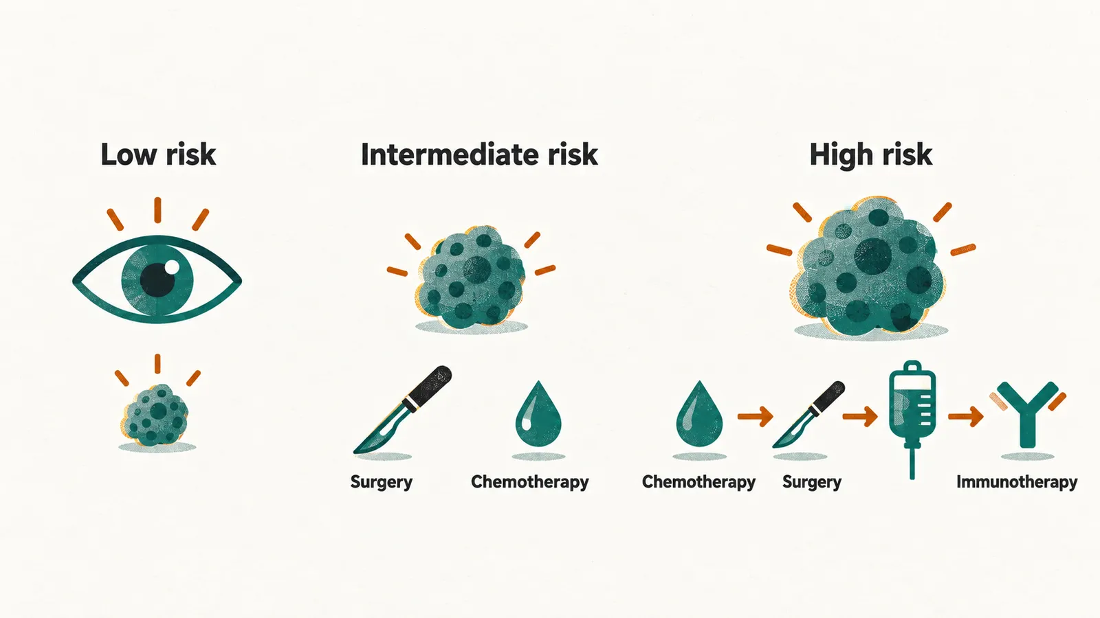
Age, stage and biology together place a child in a low-, intermediate- or high-risk group, and management is adapted to that group. The range of what is offered is unusually wide — from observation alone to the most intensive multimodal therapy in pediatrics.
- Low-risk disease is often cured by surgery alone, and selected infants with stage 4S or MS disease can be observed, because spontaneous regression is expected.
- Intermediate-risk disease is treated with surgery plus moderate-intensity chemotherapy.
- High-risk disease is treated in several steps: intensive induction chemotherapy, surgery where feasible, high-dose chemotherapy with autologous stem cell rescue (reinfusion of the child’s own previously collected stem cells), and radiotherapy to the primary site and to residual disease. Post-consolidation maintenance then combines retinoids (13-cis-retinoic acid, and all-trans retinoic acid in some regimens), to push residual neuroblasts toward differentiation, with anti-GD2 antibody immunotherapy directed at GD2 on the neuroblastoma cell surface. Dinutuximab and naxitamab are the agents in current use, supported by GM-CSF (granulocyte-macrophage colony-stimulating factor), IL-2 (interleukin-2) and isotretinoin in the regimens that established their benefit.
- Targeted radiotherapy with ¹³¹I-mIBG is used in mIBG-avid disease, particularly in the relapsed setting, and ALK inhibitors have a role in ALK-altered disease.
Prognosis
The features that set the risk group also describe the outlook. Favorable features are age under 18 months at diagnosis, a thoracic, cervical or presacral primary rather than an adrenal one, absence of lymph node involvement, hyperdiploidy, and absence of MYCN amplification. Unfavorable features are MYCN amplification, lymph node involvement, older age and high-risk image-defined disease.
Low-risk children have long-term survival of around 90%. High-risk disease remains the hard problem: despite the addition of myeloablative (high-dose) therapy, differentiation therapy and anti-GD2 immunotherapy, 5-year event-free survival in the high-risk group sits at roughly 50%.
Risk group and survival describe the course of the disease over years. Beyond that biology, the position of the tumor decides what happens to the child: a paravertebral tumor that grows into the spinal canal, or a mass that compresses the airway, presents as an emergency in its own right — one that has to be dealt with before staging and biology are settled.