
Osteoporosis causes no symptoms until it breaks a bone. The clinical picture is therefore made up of the fractures it produces and their consequences, together with the history and examination findings that point to a cause or to a high risk of falling.
Consequences of the disorder
Osteoporosis is a common systemic skeletal disorder characterised by low bone mineral density, which is the amount of mineral in the bone, and it leads to fracture, bone pain, disability and loss of independence.
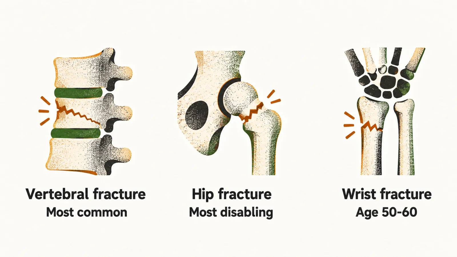
The three fracture sites that define the disorder behave differently.
Vertebral fractures are the most common osteoporotic fracture. Most are subclinical, meaning they cause no recognised symptoms; the symptomatic minority cause back pain, loss of height and kyphosis, a forward curvature of the spine. Multiple thoracic fractures can restrict the lungs, and lumbar fractures can alter the abdominal cavity enough to cause constipation, abdominal pain, early satiety and weight loss.
Hip fractures are the most disabling: about 20% of patients need long-term nursing home care, and about 60% do not fully regain their pre-fracture independence.
Wrist fractures tend to occur earlier, between 50 and 60 years of age, and are about five times more common in women; although less disabling, they predict later fractures as strongly as hip and vertebral fractures do.
A fracture in an adult aged 50 years or older should be treated as a sentinel event rather than an accident. It marks a skeleton that is likely to fracture again, particularly in the year that follows, and a previous fracture predicts the next one independently of bone mineral density.
What the medical history establishes
Because the disorder is silent until it fractures, the medical history carries much of the work of assessment. A thorough medical history in a patient with osteoporosis has three practical purposes:
- assessing the risk of future fractures
- identifying possible causes of secondary osteoporosis, the bone loss that arises from another disease, deficiency or drug
- judging whether a particular therapy is suitable — esophageal stricture, for example, is a contraindication to oral bisphosphonates
Risk of fracture and who needs testing
The history helps decide who needs a bone density test and how great the fracture risk is; the testing recommendations and risk assessment tools are set out in Diagnosis and risk assessment of osteoporosis. It also supplies the input for FRAX, the tool that turns clinical risk factors into a 10-year fracture probability: age, sex, weight, height, previous fracture, parent with hip fracture, current tobacco smoking, ever use of glucocorticoids, rheumatoid arthritis, secondary osteoporosis, alcohol intake 3 or more units per day, and if available, femoral neck bone mineral density and trabecular bone score.
Secondary causes
The history is also where the causes of secondary osteoporosis are looked for; they are set out in Secondary causes of osteoporosis.
Choosing and following treatment
Selecting the most appropriate treatment depends on identifying co-morbidities of clinical significance. For example, a high risk of breast cancer favors raloxifene use, while a history of thrombophlebitis suggests that raloxifene should not be used; esophageal stricture is a contraindication for oral bisphosphonate use; a patient with a skeletal malignancy should not be treated with teriparatide.
The history also shows the factors contributing to suboptimal response to therapy: compliance and persistence to therapy, adequacy of calcium and vitamin D, and comorbidities.
The history raises suspicions about risk, cause and suitable therapy; examining the patient and measuring bone density is how the diagnosis itself is made.