Bone mineral density in adulthood is the end result of two processes: the peak bone mass acquired during adolescence, and the loss of bone during the middle and later years. Because peak bone mass is one of the strongest determinants of fracture decades later, anything that lowers it — or that accelerates loss afterwards — lowers the density at which a person eventually fractures.
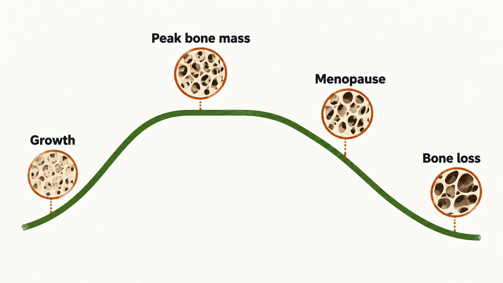
Two physiological states reduce bone mineral density sharply: the years of rapid linear growth in adolescence, when the expanding skeleton temporarily outruns its mineralisation, and the years after the menopause in women.
Peak bone mass and its determinants
Peak bone mass is the highest bone mineral density a person ever attains, and it is reached during puberty. It is higher at all skeletal sites in boys than in girls, and boys reach it about two years later; part of that male advantage reflects a larger cross-sectional bone area.
Several factors determine how high the peak is:
- nutrition, including calcium and vitamin D
- growth hormone and IGF-I (insulin-like growth factor I)
- estradiol and testosterone
- genetics
Genetics is the largest single influence: at least 50% of peak bone mass is determined by genetic factors. Because bone mineral density is a polygenic trait shaped by gene–gene and gene–environment interaction, identifying the individual genes has proved difficult.
The timing of the gonadal steroid surge matters as much as its size. The window in which bone formation is favoured lasts no more than about three years; a person who goes through puberty late therefore reaches a lower peak bone mineral density, and a lower stature, than one whose puberty is normally timed. Estradiol is required for peak density in both sexes and also closes the epiphyseal plates, the growth zones at the ends of long bones — which is why men who lack aromatase, or a functional estrogen receptor, are tall with unfused epiphyses and a very low bone mass.
Diabetes affects the skeleton, but not in the same way in both types. Type 1 diabetes, especially when it begins in childhood or adolescence, is associated with low bone mass and a higher fracture risk, because it disrupts the marrow niche and uncouples remodelling, so that formation no longer keeps pace with resorption, during the years when peak mass is being acquired. Type 2 diabetes carries roughly a 20% higher fracture risk even though areal bone mineral density is normal, and its fractures are usually peripheral — a reminder that density alone does not capture every component of bone strength.
Once peak mass has been reached, what happens to bone afterwards depends on how it is renewed, which is the work of remodelling.
The bone remodelling cycle
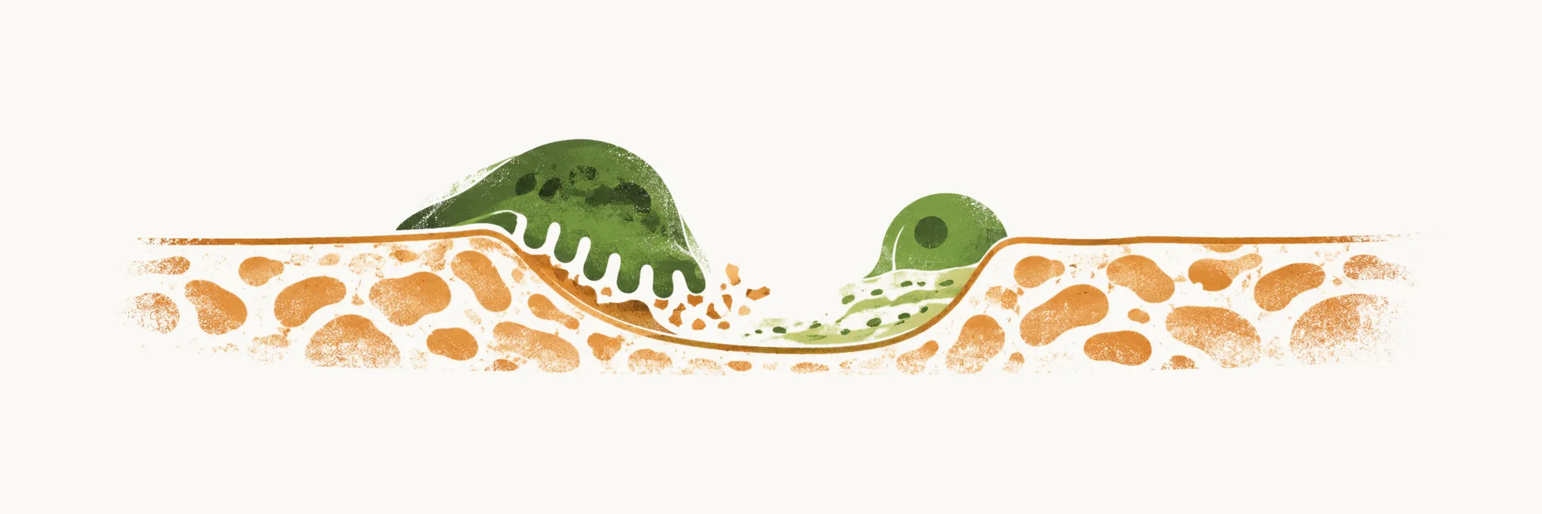
Bone remodelling is the continuous cycle in which a small volume of bone is resorbed and then replaced at the same site. It exists for two reasons: to release calcium into the extracellular fluid on demand, and to renew bone so that it keeps its elasticity and strength. Remodelling is more active in trabecular bone — the vertebrae and the proximal femur, for example — because trabecular bone has much greater surface area and lies close to the marrow, the most metabolically active compartment of the skeleton.
Coordination of the cycle rests with the osteocyte, a former osteoblast that becomes embedded in the matrix it produced. Osteocytes sense fluid shifts and microcracks — this is how the skeleton detects mechanical load and damage — and they release the signals that start a new cycle. Osteocytes and marrow stromal cells are the main source of RANKL, the signal that drives osteoclast precursors to differentiate into bone-resorbing osteoclasts. Against it stands osteoprotegerin (OPG), a soluble decoy receptor made by osteoblasts that binds RANKL and prevents it from activating the osteoclast. The RANKL–OPG balance therefore sets the rate of resorption.
Once resorption is under way, the osteoclast signals back to osteoblast precursors through Wnt and sphingosine. Osteocytes restrain the same pathway by secreting sclerostin, which blocks Wnt signalling inside the osteoblast; this restraint is what has made sclerostin a drug target. Activated osteoblasts then release IGF-I, IGF-II and TGF-β (transforming growth factor beta), which recruit further osteoblasts to the site. Growth factors deposited in the matrix during formation are stored there and released again during the next resorption, which is one way the cycle is kept coupled.
The fate of a new bone cell is decided before it forms. Mesenchymal stem cells become osteoblasts under the transcription factors RUNX2 and Osx, but the same cells become adipocytes under PPARγ. Any signal that pushes that decision toward fat therefore removes potential bone-forming cells — which is why glucocorticoid excess is accompanied both by fewer osteoblasts and by more fat in the marrow.
After osteoblasts lay down new matrix, some are enclosed by it and become osteocytes. These cells are less metabolically active, but they remain central: they are the mechanical sensors that connect load to the next round of remodelling.
Uncoupled remodelling
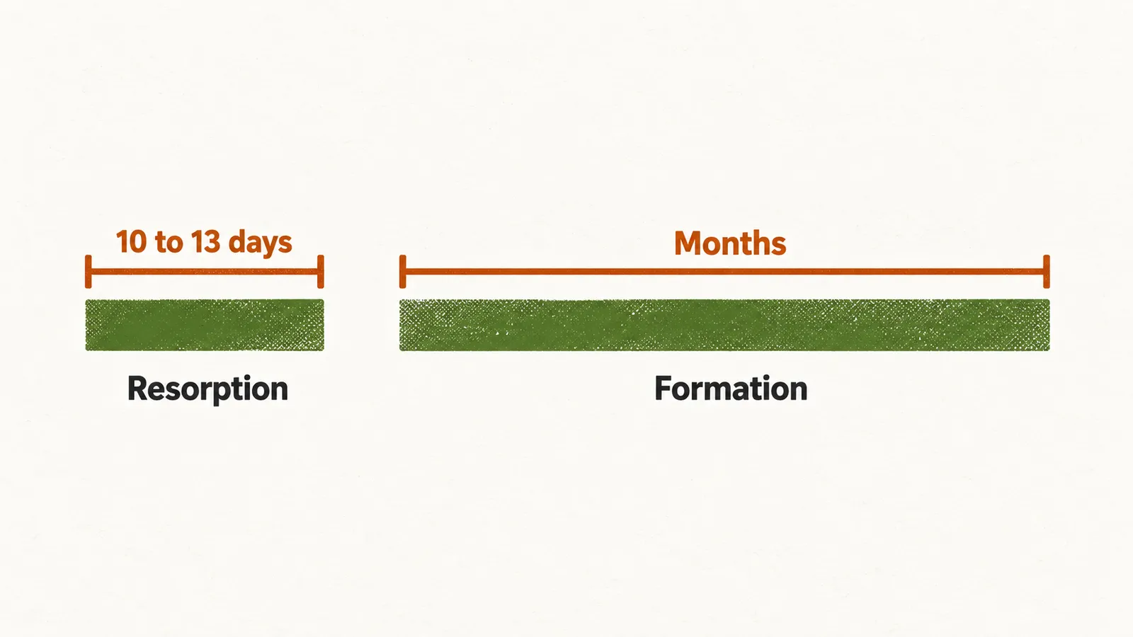
In healthy bone, resorption and formation are coupled, so the bone removed in each cycle is replaced. In osteoporosis the two are uncoupled and resorption predominates, so every cycle leaves the skeleton with slightly less bone. Part of the reason is structural: resorption takes only about 10 to 13 days, whereas formation takes months, so once the cycle accelerates, destruction outruns repair.
What tips remodelling toward resorption differs by setting, and the loss of estrogen is the most important driver of primary osteoporosis.
Estrogen loss and the postmenopausal window
Estrogen loss at the menopause is the single most important driver of primary osteoporosis, and it explains the steep bone loss of the years immediately after menopause. In a small fraction of women this loss is unusually rapid, and two signals are thought to contribute.
The first is the fall in estrogen itself. The exact molecular link is not settled. Some studies suggest that estrogen normally restrains transcription of IL-6, a cytokine (interleukin-6) that promotes resorption, so losing estrogen removes that brake. Stronger support comes from the clinic: men with an aromatase mutation or a mutated estrogen receptor, who cannot make or respond to estradiol despite normal testosterone, develop severe osteoporosis. Estradiol, not testosterone alone, is therefore the key hormone for the skeleton in both sexes.
The second is follicle-stimulating hormone (FSH). In a mouse model, a high FSH level was associated with greater bone loss independently of estrogen, although this has not been confirmed in humans.
Vitamin D deficiency and secondary hyperparathyroidism in the elderly
Remodelling also responds to the calcium supply. Among people in their 80s and 90s, and especially women, a serum 25-hydroxyvitamin D level below 20 ng/mL leads to secondary hyperparathyroidism, a raised parathyroid hormone (PTH) level. The low calcium supply drives PTH up, and PTH stimulates the osteoblasts and provokes remodelling. Because bone formation in the elderly does not rise in step with resorption, the cycle becomes further uncoupled and bone loss accelerates.
Glucocorticoid excess
Glucocorticoids are the second most common cause of low bone mass in the general population and one of the most common causes of osteoporotic fracture. They act through several routes at once, some indirect, through parathyroid and gonadal hormones and calcium handling, and some directly on bone cells, which is why bone loss begins within the first few months of therapy and is both rapid and sustained. In the table, LH is luteinizing hormone and 1,25 D is 1,25-dihydroxyvitamin D.
| Response to glucocorticoids | Effects on bone remodelling | Effects on bone mass |
|---|---|---|
| Increased PTH secretion | Increased bone resorption? decreased bone formation | rapid loss of bone |
| Decreased LH/FSH secretion | Increased bone resorption due to loss of estrogen | loss of bone |
| Impaired calcium absorption due to decreased 1,25 D | Increased PTH, increased bone resorption | loss of bone |
| Increased calcium loss in urine | Secondary increase in PTH, increased bone resorption | loss of bone |
| Acute suppression of osteoblasts and apoptosis | Reduced bone formation | gradual bone loss |
| Stimulation of osteoclastogenesis | Increased bone resorption | rapid loss of bone |
However it arises, this loss of bone gives no symptoms of its own, so the disorder usually first shows itself through what it does to the skeleton.