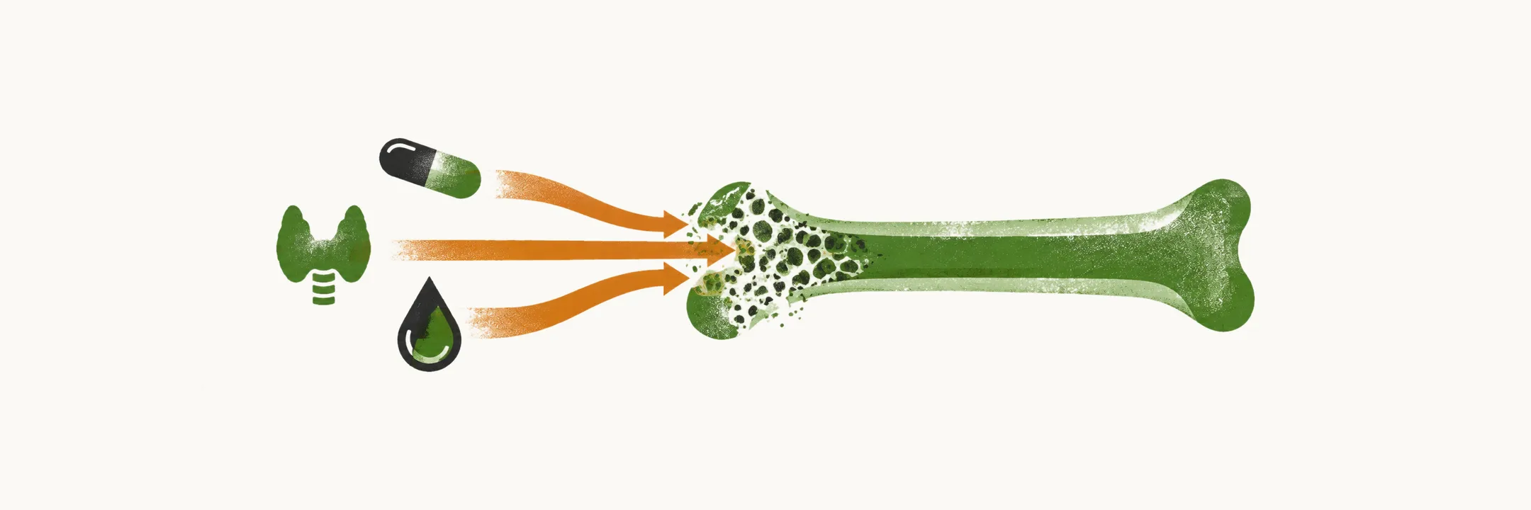
Most osteoporosis is primary: it follows from ageing and from the loss of estrogen after the menopause, and no separate cause needs to be found. In a substantial minority of patients, however, bone loss is driven by another disease, a deficiency or a drug. Identifying that cause matters, because treating it can stop the bone loss — and because some causes, such as multiple myeloma, carry consequences of their own.
Which conditions cause secondary osteoporosis
Secondary causes fall into groups, from lifestyle and inherited disorders to endocrine, digestive, blood, rheumatic and neurological disease and drugs:
| Group | Causes |
|---|---|
| Lifestyle | Low calcium intake, vitamin D deficiency, alcohol excess, excess vitamin A, high salt intake, inadequate physical activity, frequent falling |
| Genetic | Osteogenesis imperfecta, Marfan syndrome, homocystinuria, hypophosphatasia, cystic fibrosis, idiopathic hypercalciuria, glycogen storage diseases, Riley-Day syndrome, Menkes steely hair syndrome, androgen insensitivity |
| Endocrine | Hypogonadism, hyperthyroidism, hyperparathyroidism, Cushing’s syndrome, panhypopituitarism, anorexia nervosa, premature menopause, obesity |
| Gastrointestinal and hepatic | Celiac disease, malabsorption, chronic liver disease, primary biliary cirrhosis, pancreatic disease |
| Hematologic | Multiple myeloma, lymphoma, systemic mastocytosis, thalassemia, hemophilia |
| Rheumatic, neurological and other chronic disease | Rheumatoid arthritis, ankylosing spondylitis, muscular dystrophy, spinal cord injury, immobilisation, epilepsy, chronic obstructive pulmonary disease, congestive heart failure, parenteral nutrition, hyponatremia, amyloidosis, a previous fracture in adulthood, HIV/AIDS |
| Medications | Glucocorticoids (>5 mg of prednisone or equivalent for >3 months), anticonvulsants, long-term heparin, excess thyroid hormone, GnRH agonists, aromatase inhibitors, lithium, depo-medroxyprogesterone, tacrolimus, proton pump inhibitors, selective serotonin reuptake inhibitors |
A few of these deserve more than their place in the table. Glucocorticoid excess is the second most common cause of low bone mass in the general population, and because it acts through several mechanisms at once, bone loss begins within the first few months of treatment. Endocrine causes are common and often treatable: hyperthyroidism raises bone turnover, hyperparathyroidism increases resorption, and Cushing’s syndrome produces the same effect as therapeutic glucocorticoids. Malabsorption and celiac disease are easy to miss, because they lower the absorption of calcium and vitamin D without obvious gastrointestinal symptoms. Multiple myeloma and systemic mastocytosis can produce vertebral fractures that look osteoporotic, which is why myeloma enters the differential of any patient presenting with them.
The pattern in men differs from that in women. The commonest secondary causes in men are glucocorticoid excess, hypogonadism and alcohol excess, and a proportion of men have idiopathic osteoporosis — low bone formation with low IGF-I — for which no cause is found.
How a secondary cause is suspected
A Z-score, the number of standard deviations by which bone mineral density differs from the mean for the patient’s own age and sex, is sometimes used as a prompt to look for a secondary cause when it lies more than 2 standard deviations below that mean, although this criterion is not universally accepted. Clinical clues point the same way: an enlarged thyroid gland, atrophic testicles, or the combination of blue sclera, yellow-brown teeth and hearing loss all suggest a specific cause.
A short list of laboratory tests is then used to screen for the treatable causes. ESR is the erythrocyte sedimentation rate, TSH is thyroid-stimulating hormone, and anti-EMA and anti-tTG are antibodies against endomysium and tissue transglutaminase:
| Test | Abnormal finding | Suggests |
|---|---|---|
| Complete blood count, ESR | Anemia | Malnutrition |
| Raised ESR | Multiple myeloma | |
| Serum calcium | Hypocalcemia | Vitamin D deficiency |
| Hypercalcemia | Vitamin D intoxication, primary or secondary hyperparathyroidism, thyrotoxicosis, renal failure, Paget’s disease | |
| Serum phosphate | Hypophosphatemia | Primary or secondary hyperparathyroidism, vitamin D deficiency |
| Alkaline phosphatase | Raised | Healing fractures, Paget’s disease, osteomalacia |
| 25-OH vitamin D | Low | Low vitamin D stores; values of 30-50 ng/ml are often taken as optimal, though there is no consensus |
| Creatinine | Raised | Chronic kidney disease, a common cause of secondary hyperparathyroidism and skeletal disease |
| TSH | Low | Hyperthyroidism, which increases bone turnover |
| Liver enzymes | Raised | Chronic liver disease |
| Urinalysis | Proteinuria | Multiple myeloma or chronic kidney disease |
| 24-hour urine calcium | Hypercalciuria | A renal leak of calcium (renal), excess skeletal loss (resorptive), or high gut absorption (absorptive) |
| Anti-EMA and anti-tTG | Positive | Celiac disease |
| Serum protein electrophoresis and free kappa/lambda ratio | M component | Multiple myeloma |
| Dexamethasone suppression test | Failure to suppress | Cushing’s syndrome |
| Testosterone (total and free) | Low | Hypogonadism in men |
Finding a secondary cause does not always remove the need for treatment of the skeleton itself. Correcting the cause may reduce fracture risk, but the patient can still be left with bone loss, and some causes — such as continued glucocorticoid therapy — have to be managed while the drug is still required.