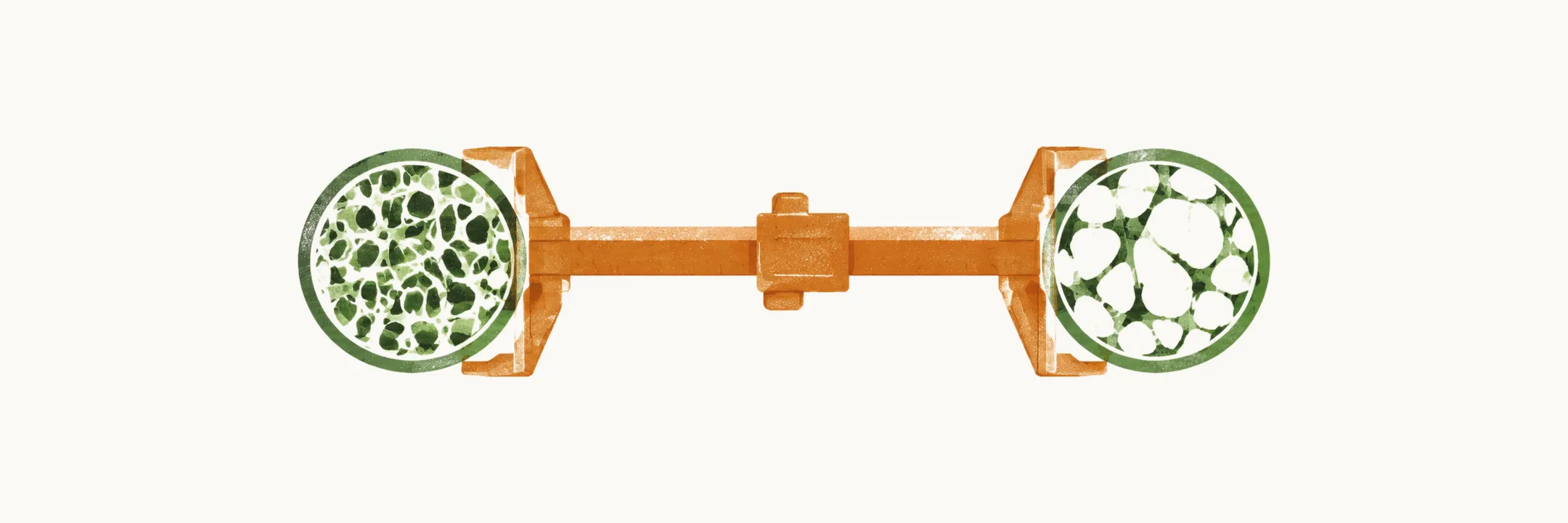
Osteoporosis is a disorder of reduced bone strength, and whether a person with it will fracture depends on more than one factor. Which factors matter most differs by skeletal site; among them, low bone mineral density is the strongest single predictor of hip and spine fractures.
Bone mineral density
Bone mineral density (BMD) is a quantitative measure of the mineral in bone, usually measured by dual-energy X-ray absorptiometry (DXA). Its relationship to fracture is continuous: each standard deviation below the young-adult mean at almost any skeletal site is associated with a nearly two-fold higher risk of hip fracture. That relationship holds all the way below the mean, without a threshold, so a density measurement is informative even in someone who has not yet fractured.
Other risk factors
The risk factors for hip fracture in particular are:
- age over 65 years
- a history of spine or hip fracture
- a maternal history of hip fracture
- weight loss after the age of 50
- low body mass index
- poor neuromuscular function
Clinical risk factors that act on fracture risk independently of density include older age, any previous fracture, a higher number of previous fractures, more severe previous fractures, and the conditions that make falling more likely.
Measurable and unmeasurable determinants of bone strength
Density is not the whole of bone strength. Not every determinant of bone strength can be measured, and not every measurable one is a proven risk factor. In the table, areal BMD is density per area of bone as DXA reports it, volumetric BMD is density per volume as quantitative computed tomography (QCT) reports it, pQCT is peripheral QCT, TBS is the trabecular bone score, a measure of trabecular connectivity derived from the DXA image, and VFA is vertebral fracture assessment. In the middle column, +/- and ? mark factors whose status as a risk factor is uncertain.
| Characteristic | Recognised risk factor for fracture | Clinically measurable |
|---|---|---|
| Bone Mineral Density (areal) | Yes | Yes (DXA) |
| Bone Mineral Density (volumetric) | Yes | Yes (QCT) |
| Microcracks | No | No (histology) |
| Trabecular Connectivity | Yes | TBS (DXA) |
| Periosteal Circumference | No | Yes (pQCT/DXA) |
| Cortical Thickness | +/- | Yes (pQCT/DXA) |
| Bone Turnover | Yes | Yes (markers) |
| Previous Osteoporotic Fracture | Yes | Yes (radiographs or VFA) |
| Mineralization and cortical porosity | ? | No/yes (back scatter EM) and sometimes with high resolution microCT |
Putting the pieces together: FRAX
The best tool for putting these pieces together is FRAX, which combines femoral neck bone mineral density with clinical risk factors — previous fracture, glucocorticoid intake, alcohol consumption and others — to give a 10-year probability of hip fracture and of major osteoporotic fracture. Density remains FRAX’s main predictor, because it is quantitative. What the tool cannot weigh fully are the qualitative properties of bone:
- bone turnover rate
- trabecular bone connectivity
- cortical and periosteal bone size
- skeletal morphometry
Trabecular connectivity shows why density alone is not enough. Two people can have the same bone mineral density and still differ in risk, because the one whose trabecular network is more disrupted has less of the internal architecture that carries load. Which processes change density, turnover and architecture in the first place is the subject of bone physiology.