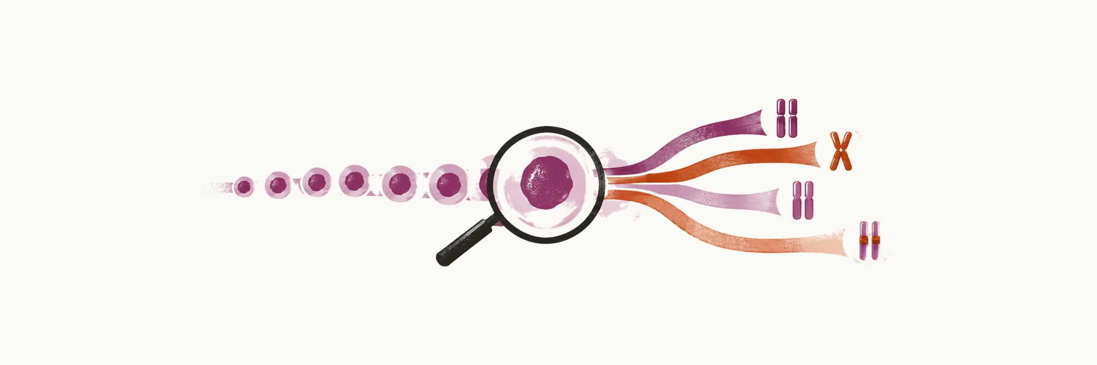
The classification of acute myeloid leukemia has moved from what the blasts look like to what their chromosomes and genes carry, and the categories the WHO defines are the ones now used to name the disease.
How AML is classified
The most recent classification of AML is made by the WHO, and it is based mainly on cytogenetic (chromosomal) and molecular abnormalities, morphology (how the cells look under the microscope) and clinical features. An earlier classification, proposed by the French-American-British (FAB) council, was based on morphology alone. Genetic studies are now one of the most efficient ways of classifying AML and of deciding which subtype a patient has.
The blast count
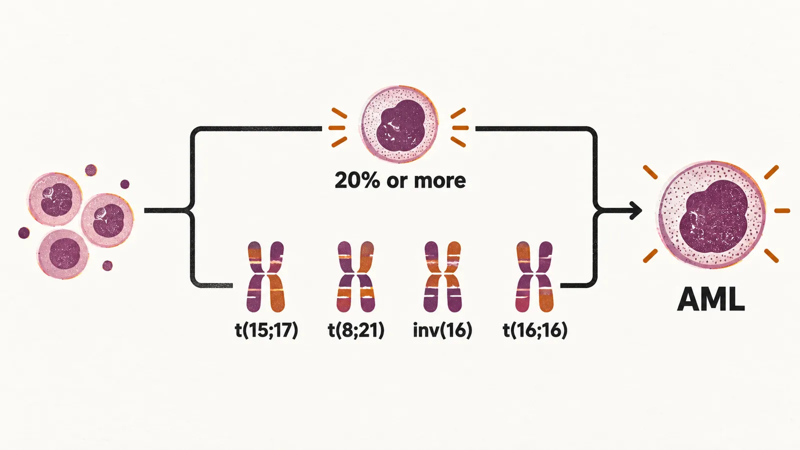
A marrow or blood blast count of 20% or more is needed for the diagnosis of AML. There are defined exceptions: AML with one of the recurrent genetic abnormalities t(15;17), t(8;21), inv(16) or t(16;16) is diagnosed even when the blast count is lower, because those abnormalities are specific enough to define the disease on their own.
The WHO categories
Based on the WHO classification, AML can broadly be placed in one of the groups below:
- AML with recurrent genetic abnormalities
- AML with myelodysplasia-related changes, including cases without a history of prior cytotoxic therapy
- therapy-related myeloid neoplasm
- AML not otherwise specified
- myeloid sarcoma
Recurrent genetic abnormalities
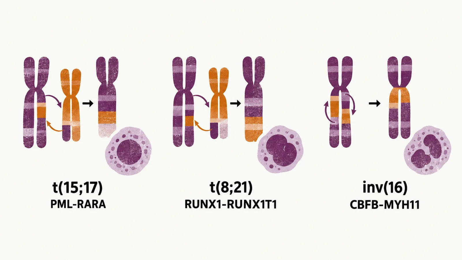
A few named genetic findings define the most important subtypes. Acute promyelocytic leukemia (APL) carries t(15;17), which forms the PML-RARA fusion product, a hybrid gene made when a translocation joins two genes. Core binding factor (CBF) AML carries t(8;21), which forms the RUNX1-RUNX1T1 fusion product, or inv(16) and t(16;16), which form the CBFB-MYH11 fusion product. Both groups of fusion proteins act by disrupting the transcription factors that cells need to mature, which is why the blasts do not differentiate.
Some forms of AML have characteristic morphological features that point to the underlying abnormality:
- AML with myelodysplasia-related changes, which shows dysplasia in the blood and marrow
- APL with t(15;17) or the PML-RARA fusion
- AML with inv(16), which has abnormal bone marrow eosinophils
- AML with t(8;21), which shows increased CD19, slender Auer rods, and an increased number of morphologically normal eosinophils
- AML with a mutation of nucleophosmin (NPM1) together with fms-related tyrosine kinase 3 (FLT3), which shows a cup-shaped nucleus
Immunophenotype
Beside the morphological and genetic approach, immunophenotyping, which identifies the proteins (biomarkers) that cells express, with the help of flow cytometry is sometimes needed to assign a lineage when the blasts are too immature to classify morphologically. AML with minimal differentiation is characterised by undifferentiated cells that express the myeloid lineage biomarkers CD13 and CD117, and acute megakaryoblastic leukemia can be identified with platelet-associated biomarkers such as CD41 and CD61.
Other findings that carry information
Some abnormalities are not a category of their own but change how the disease behaves. The FLT3-ITD mutation, which has a bad prognostic impact, mostly occurs in patients with cytogenetically normal AML (no visible chromosomal abnormality), or CN-AML. Age changes the pattern: in younger cases of AML, t(8;21) and t(15;17) are seen more commonly, while at older ages the TP53 mutation, del(5) and del(7) are more common.
Trisomies (an extra copy of a chromosome) make individuals more prone to AML and ALL, in particular trisomy of chromosomes 8 and 21. Beside chromosomal aberrations, some AML are defined by mutations such as mutated NPM1 or mutated CEBPA.
Two further associations point to how the disease presents: DIC (disseminated intravascular coagulation) related to AML is mostly related to t(15;17), and myeloid sarcoma, a tumor mass of myeloid blasts, is mostly related to t(8;21).