
The diagnosis of acute myeloid leukemia rests on finding blast cells in the peripheral blood or the bone marrow, and on the laboratory work-up that shows what kind of blasts they are.
When to suspect AML
AML should be considered in any patient who has circulating blast cells, unexplained cytopenia (a low count of one or more blood cell types) with or without related symptoms, or an unexplained metabolic or oncologic emergency such as tumor lysis syndrome or hyperleukocytosis (a very high white cell count).
Diagnostic criteria
The diagnosis of AML is based on infiltration of the peripheral blood or bone marrow by 20% or more blast cells, which show characteristic morphologic, cytochemical and immunophenotypical features. In some cases, even when fewer than 20% of the typical blast cells of AML are present, the patient can be diagnosed with AML if there are myeloid sarcoma manifestations or one of the definitive cytogenetic abnormalities of AML: inv(16), t(16;16), t(8;21) or t(15;17). The lower threshold exists because these findings are, on their own, enough to establish the disease.
The laboratory work-up
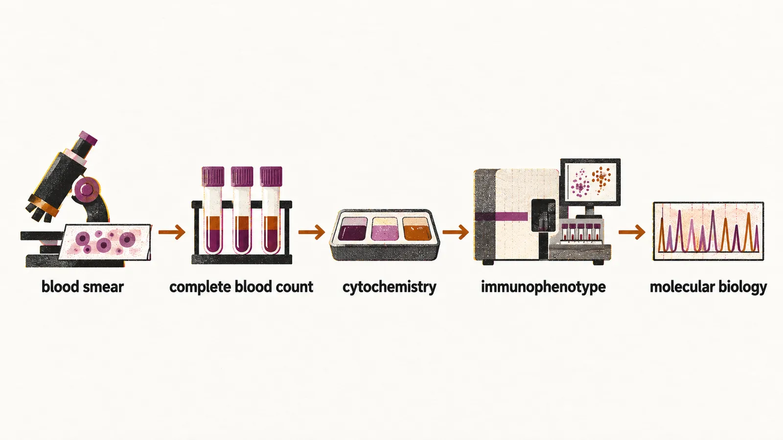
The diagnosis of a patient suspected of having acute leukemia should be made through morphology of the blood cells in the peripheral blood smear, a complete blood count, LDH and uric acid (both very high in acute leukemia), cytohistochemistry, immunophenotype, and molecular biology, which is the gold standard way of diagnosis. In acute promyelocytic leukemia, a very rapid diagnosis of the subtype is needed because of the hemorrhagic complication of DIC (disseminated intravascular coagulation).
Morphology and immunophenotype
The blood and marrow films give the first and often the decisive evidence. Abnormal rod-shaped granules called Auer rods are not present in all cases, but if they are present the diagnosis of AML is certain.
Cytochemistry helps when the morphology alone is not enough. On staining, the blasts are positive for Sudan black in M1 to M4 (subtypes of the earlier FAB classification), because of the presence of peroxidase, and show dual esterase in M4 and M5, because of the presence of acid-alpha naphthyl acetate esterase (ANAE).
For immunophenotyping, MPO (myeloperoxidase), CD33 and CD13 can be used. The markers that identify the lineage in the less common forms are:
- monocytic: CD14
- myelocyte: CD15
- megakaryocytic leukemia: CD41 and CD61
- erythroid leukemia: glycophorin A
- NK leukemia: CD16 and CD56
Differential diagnosis
The important differential diagnoses of AML are infection, aplastic anemia, MDS/MPN (myelodysplastic/myeloproliferative neoplasms) such as CMML (chronic myelomonocytic leukemia) — in which the differentiation from AML is based on the percentage of blast cells in the bone marrow — and CML (chronic myeloid leukemia) with acute blastic crisis.