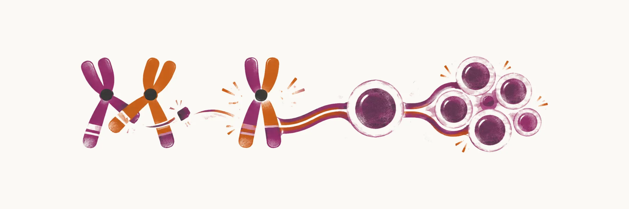
Acute myeloid leukemia begins in a single hematopoietic stem or progenitor cell. That cell acquires somatic mutations that give it and its descendants a growth and survival advantage, and the resulting clone expands while accumulating further mutations. Normal hematopoiesis depends on orderly maturation, and the leukemic clone cannot mature, so the marrow gradually fills with non-functional precursor cells while the production of normal blood cells falls.
The types of genetic lesion
The genetic changes behind acute leukemia appear mostly in the form of translocations (segments exchanged between chromosomes), mutations, deletions (loss of a chromosome segment), hyperdiploidy or hypodiploidy (more or fewer chromosomes than normal). At the chromosome level, balanced translocations are the most common abnormality in acute leukemia, at about 40%. The remaining lesions are mostly unbalanced, and fall into deletions, trisomies (an extra copy of a chromosome) and hyperdiploidy, and hypodiploidy. At the gene level, the effect is usually the activation of an oncogene (a gene that promotes growth) or the loss of a tumor suppressor gene (a gene that restrains growth). These lesions differ in what they do to the cell, and the mutations responsible can be sorted into two functional classes.
Two classes of mutation cooperate
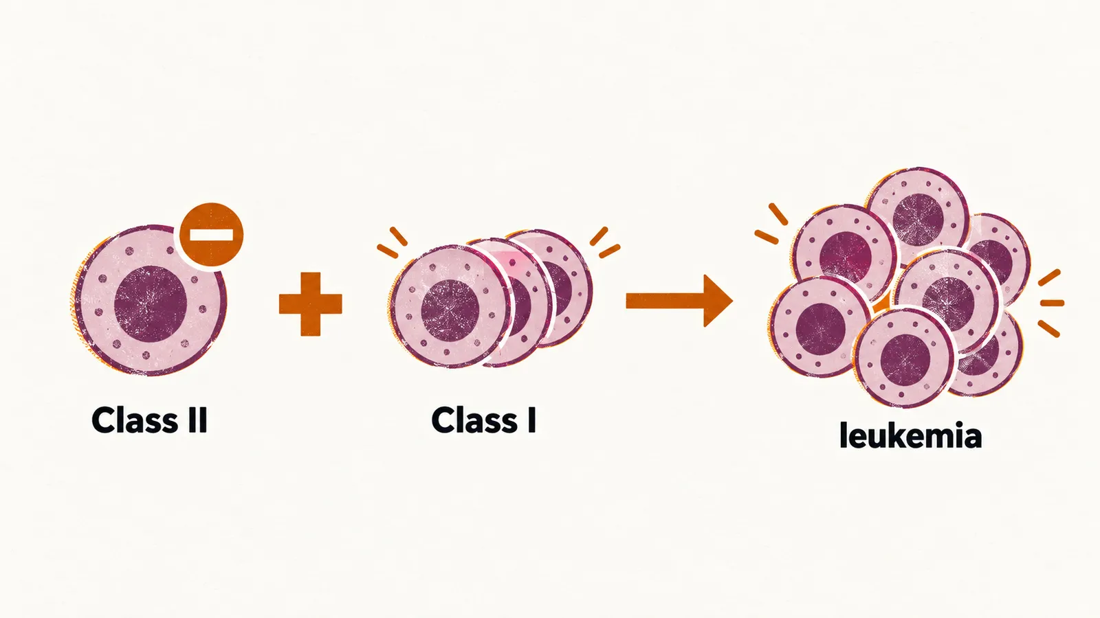
No single mutation is usually enough. The two-hit model of leukemogenesis divides the responsible mutations into two functional classes. Class II mutations block myeloid differentiation, so the cells stay immature; they include the fusion genes formed by t(8;21) (RUNX1-RUNX1T1), inv(16) (CBFB-MYH11) and t(15;17) (PML-RARA), and mutations in transcription factors such as CEBPA and RUNX1. Class I mutations drive proliferation and survival, and include FLT3, NRAS and KIT. A differentiation block alone leaves cells abnormal but not aggressively expanding; a proliferative mutation alone does not stop the cells maturing. When both are present in the same clone, the cells multiply without maturing, and leukemia develops. The model is a useful simplification: sequencing now shows that most AML genomes carry several driver mutations, and the order in which they are acquired shapes the disease.
Clonal evolution and the aging marrow
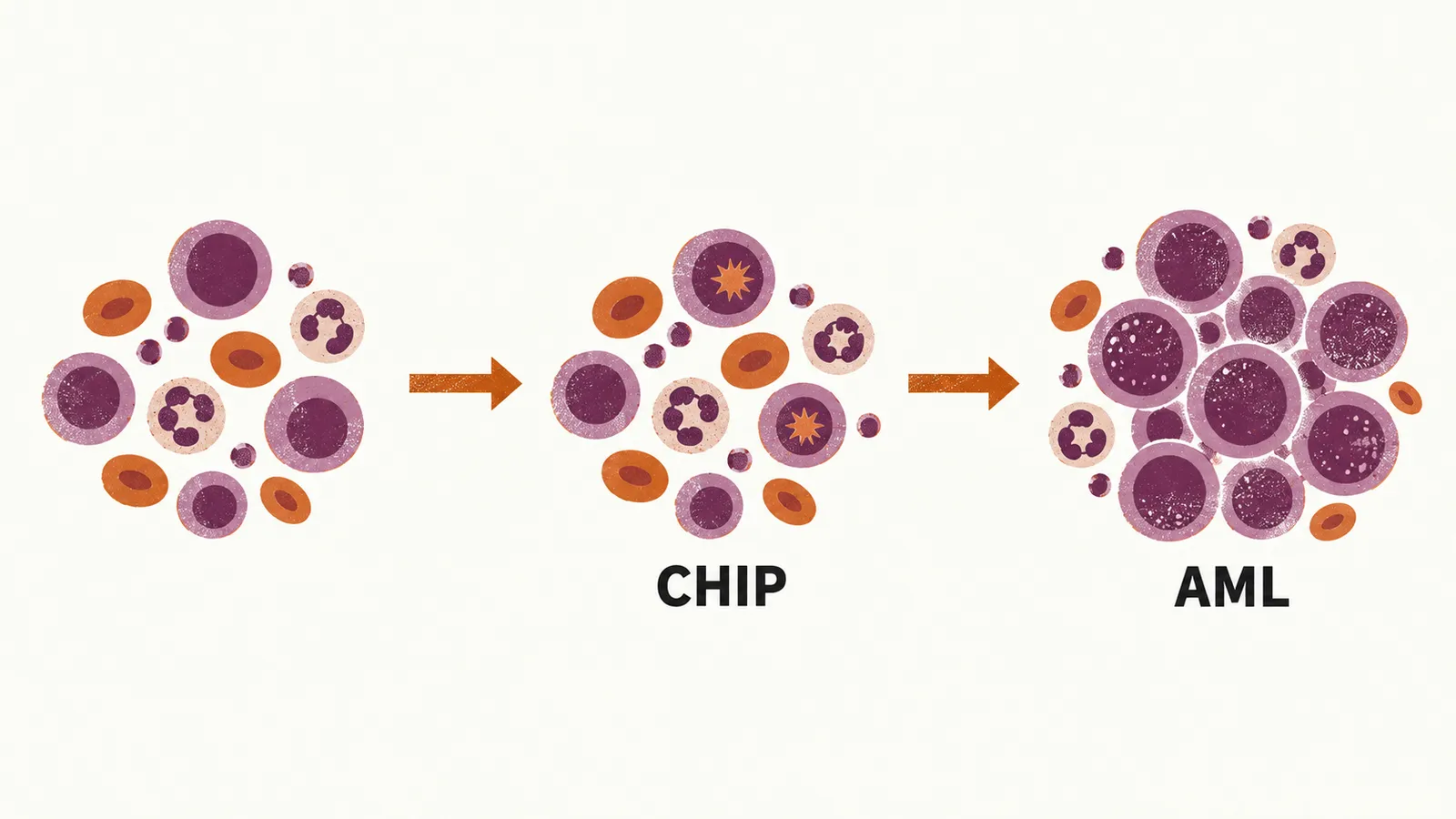
Genome sequencing studies show that AML arises from the accumulation of mutations during the process of aging, and that the mutations accumulate in the same clone over time. An early and common stage is detectable in people who are well: in the peripheral blood of 5-7% of individuals over 70 years old there are mutated, clonally expanded cells without overt disease. Calling these cells pre-malignant is not accurate; it is better to say clonal hematopoiesis of indeterminate potential (CHIP), which is mostly related to genomic lesions in epigenetic modifiers such as DNMT3A, TET2 and ASXL1. CHIP is a precursor state rather than a disease: most people who have it never develop leukemia, but it raises the risk, and further mutations acquired by the same clone are what convert it into AML. The aging marrow environment favours the process, which helps explain why the disease is concentrated in older adults.
Primary, secondary and therapy-related disease
Not every AML begins in a healthy marrow. Some cases arise from an existing myeloid disorder, or after cytotoxic treatment for an earlier cancer, when a clone that is already present — in a myelodysplastic marrow, or left behind by chemotherapy or radiotherapy — acquires the additional hits that make it acute. This is why AML with myelodysplasia-related changes and therapy-related myeloid neoplasm are recognized as distinct categories.
From clone to marrow failure
As the clone expands it crowds the marrow and suppresses normal hematopoiesis, so red cells, platelets and neutrophils are produced in falling numbers. This is the source of the anemia, bleeding and infection that bring most patients to medical attention. The blasts also spill out of the marrow into the blood and infiltrate other tissues, which is why the disease can present as an organ mass or with infiltration of the gums, skin or meninges.