
Lymphocytosis means a higher than normal number of lymphocytes in the blood, and it is the finding that usually brings chronic lymphocytic leukemia (CLL) to attention. Understanding it starts with what the circulating lymphocytes are, because the count alone does not say which subset has risen or why.
Lymphocyte subsets in the circulation
About 90% of the lymphocytes in the circulation are cells of adaptive immunity, which are B and T cells, and about 10% of the lymphocytes belong to the lineage of innate immunity, which are the natural killer (NK) cells. The three lineages can be told apart by a few surface markers: CD3+ cells are T cells, CD19+ or CD20+ cells are B cells, and CD3- cells that are CD56+ are NK cells. Because CD56 identifies NK cells only once CD3 has excluded the T cells, the panel separates all three lineages at once, and the CD19+ population is the one that matters when a B-cell leukemia is suspected.
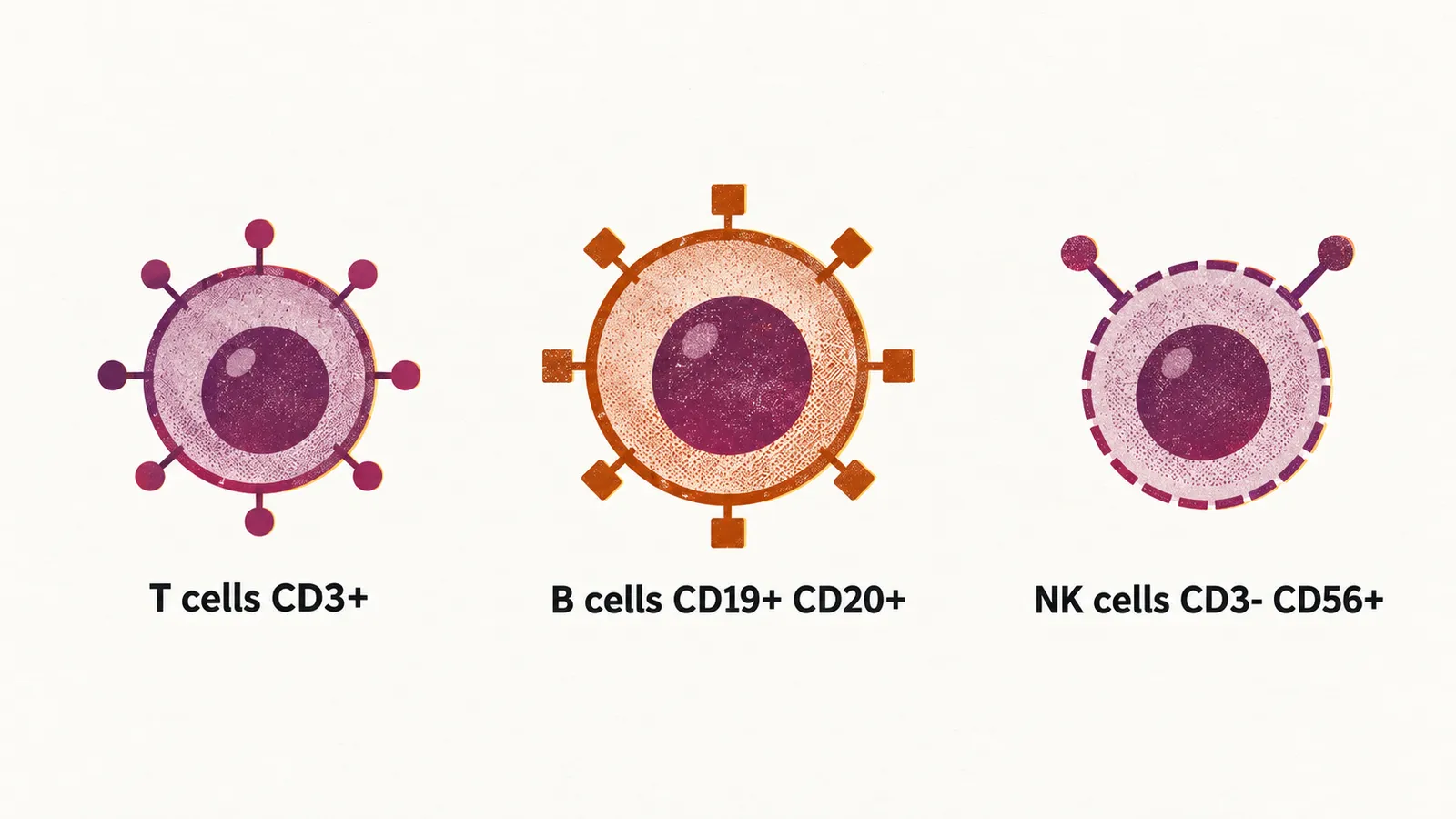
Reactive causes of lymphocytosis
In CLL the very first thing that can be seen is lymphocytosis, but lymphocytosis can be due to several hematological conditions and also to reactive processes, and these have to be distinguished from one another. Reactive lymphocytosis follows infection, whether bacterial, as with Bordetella pertussis, viral, as with cytomegalovirus (CMV) and Epstein-Barr virus (EBV), or parasitic. It also follows trauma or other acute stress such as a cardiac event or a seizure, and appears after splenectomy, in hyperthyroidism, and with a range of drugs.
Findings that point toward leukemia
Because lymphocytosis can be just a reactive response of the body to an infectious or non-infectious cause, leukemia is not what has to be considered all of the time. It becomes more likely when the rest of the picture points that way. On the peripheral blood smear, the findings that raise suspicion are cytopenia in another lineage, that is, a low count of another blood cell type, such as anemia; atypical lymphocyte morphology, although abnormally shaped lymphocytes also appear in infectious mononucleosis, so morphology alone is not proof; immature cells; concurrent hemolysis, the destruction of red cells; and a lymphocytosis that is rising rapidly. On examination, hepatosplenomegaly (an enlarged liver and spleen) and lymphadenopathy (enlarged lymph nodes) shift the balance toward leukemia. The level of the count matters as well, because there is a meaningful difference between a 50% increase in the number of lymphocytes and a 5-fold increase in their number.
Confirming clonality
Clonality, a population derived from a single cell, is the defining characteristic of leukemia, so when it is considered it has to be confirmed in the cells of the patient. Flow cytometry, which reads the surface markers of individual cells, is the first step: it shows an altered kappa:lambda ratio, meaning that the cells are restricted to one light chain (kappa or lambda, the two types of immunoglobulin light chain), and it can show abnormal markers, such as co-expression of CD5 on the B cells, or the absence of markers that normal B cells carry. Molecular studies add the presence of a clonal B-cell receptor (BCR) rearrangement and, where useful, analysis of somatic hypermutation. Together these tests separate a true clone from a reactive expansion. Whether a confirmed clone already amounts to leukemia then depends on how large it is and what else accompanies it.