
A clonal population of B cells is not automatically a cancer. The earliest detectable step of the sequence that ends in chronic lymphocytic leukemia (CLL) is monoclonal B-cell lymphocytosis, a clone that is too small to meet the criteria for leukemia.
Not every clonal proliferation is treated as cancer
A very important consideration in hematology is that not all clonality is considered an aggressive cancer or treated as cancer. In monoclonal gammopathy of undetermined significance (MGUS), for instance, there is a clonal proliferation that is not even treated in many cases. The reason is that carcinogenesis generally works by the accumulation of mutations: a detectable clone, and even a sign such as lymphocytosis in the blood, can appear long before the process produces the aggressive form of the disease. Those intermediate states need their own names and thresholds, so that a clone which is not yet leukemia is not treated as though it were.
The sequence from monoclonal B-cell lymphocytosis to Richter transformation
The evolution of malignancy for B cells follows a chronological sequence, in which chronic lymphocytic leukemia (CLL) is present in an asymptomatic and then a symptomatic phase, and Richter transformation is the development of an aggressive large-cell lymphoma from it:
- monoclonal B-cell lymphocytosis, or MBL
- asymptomatic CLL
- symptomatic CLL
- Richter transformation
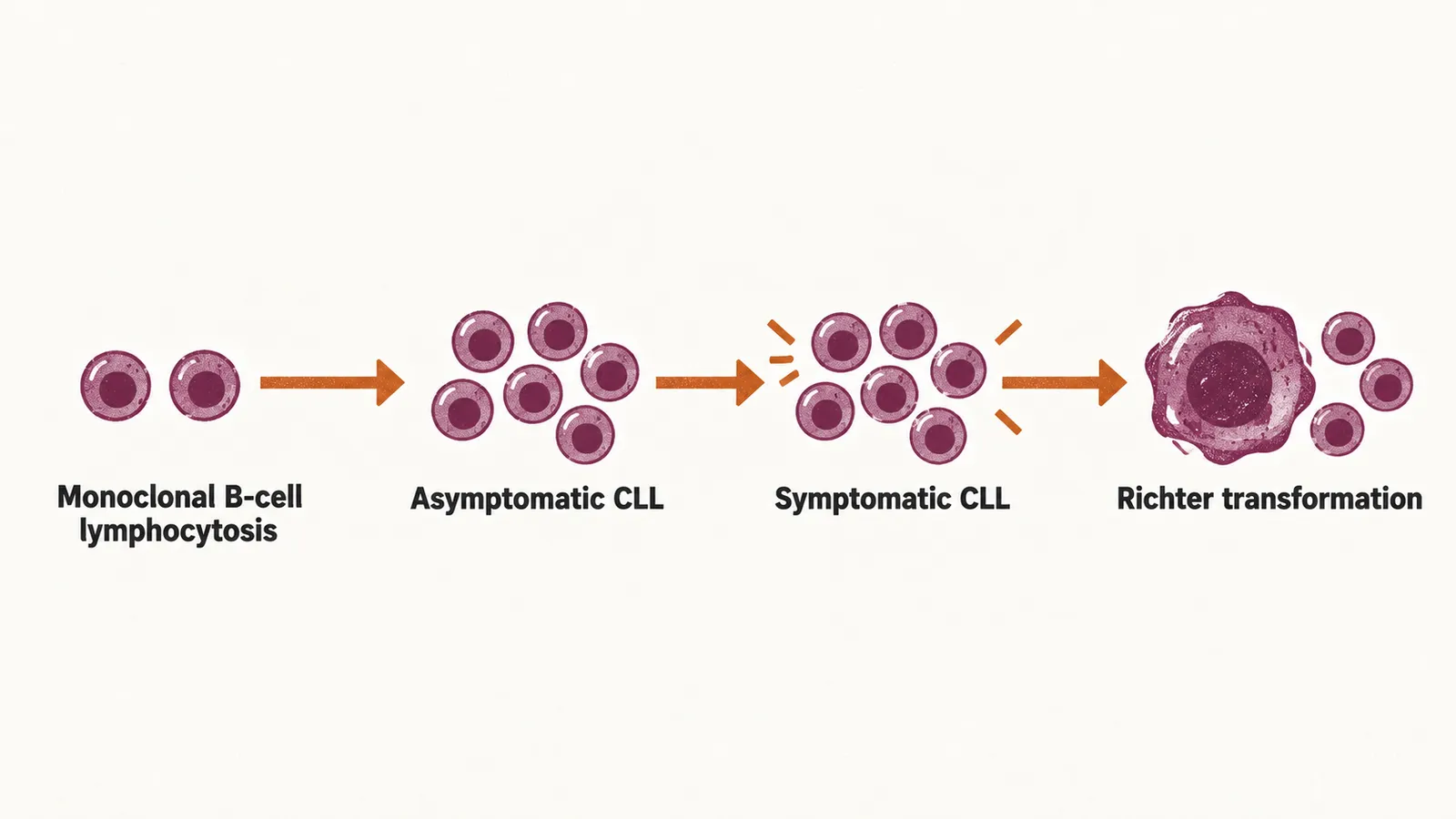
What monoclonal B-cell lymphocytosis is
Monoclonal B-cell lymphocytosis, or MBL, is defined by two conditions together: fewer than 5000 B cells per microliter, persisting for at least 3 months of follow-up; and the absence of other signs of a B-cell lymphoproliferative disorder, meaning a disease in which lymphocytes multiply abnormally. Those other signs are lymphadenopathy (enlarged lymph nodes); hepatomegaly and/or splenomegaly (an enlarged liver and/or spleen); cytopenia in another lineage; and extramedullary involvement, that is, disease outside the bone marrow. A patient who has the clonal cells and also one of these signs does not have MBL.
How often MBL is found
MBL becomes more common with age: it is found in less than 1% of the population under 40 years old and in more than 10% of the population over 70 years old. That frequency means a small clonal B-cell population is a common incidental finding in older people rather than a rare one.
The two forms of MBL
MBL is divided by the size of the clone, and the distribution of MBL is bimodal, meaning that it has two peaks, so the two forms behave differently:
| Form | Definition | Behaviour in practice |
|---|---|---|
| Low-count MBL | fewer than 500 clonal B cells per microliter | the less frequent form in clinical practice |
| High-count MBL | at least 500 clonal B cells per microliter | the more frequent form in clinical practice; the risk of developing CLL is 1% to 2% per year |
That risk in high-count MBL leads to the questions of what the clone is, why it accumulates and which features shape how it behaves, which are the questions of the disease’s biology.