
The chronic lymphocytic leukemia (CLL) cell is a mature B lymphocyte that should have died but does not. Its slow accumulation, rather than rapid division, is what produces the disease, and the same features that let it survive also give it a recognisable surface pattern and a set of recurring genetic lesions.
What the CLL cell is
The malignant cell is a mature B lymphocyte that has already met its antigen (antigen-experienced). Its numbers grow less because it divides quickly than because it escapes programmed cell death: the cells over-express BCL2, an anti-apoptotic protein, so the apoptosis that would normally remove aged lymphocytes does not happen. Division still occurs, but it is concentrated in the lymph nodes and bone marrow, in small clusters called proliferation centres, where the cells receive survival and growth signals from companion T cells, stromal cells and nurse-like cells. The B-cell receptor (BCR) on the surface of the CLL cell relays much of that signalling through kinases, enzymes that switch other proteins on, such as Bruton tyrosine kinase (BTK) and phosphatidylinositol 3-kinase (PI3K). This is why the disease is treated by blocking the BCR pathway or by restoring the apoptosis that BCL2 blocks.
The immunophenotype that identifies it
The clone is identified by the combination of markers its cells carry, read on flow cytometry. CLL cells are positive for CD19 and CD23 and co-express CD5, a marker otherwise associated mainly with T cells, while CD20 and surface immunoglobulin are only weakly expressed. The surface immunoglobulin is restricted to a single light chain, kappa or lambda, which is what shows that the population is monoclonal. The cells also express CD200 and bright CD43 and are negative for cyclin D1, a pattern that separates CLL from mantle cell lymphoma, the other CD5-positive B-cell lymphoma. CLL lymphocytes are unusually fragile, so many of them rupture during blood film preparation and are seen as smudge cells.
The cytogenetic abnormalities behind the behaviour
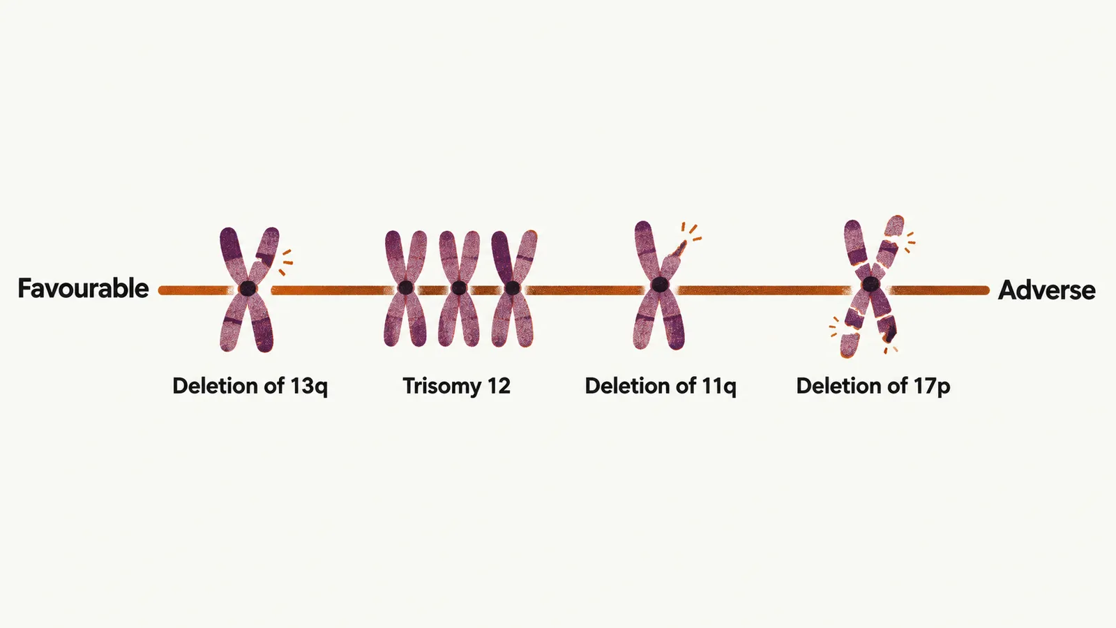
Recurring chromosome abnormalities are present in most patients and shape how the disease behaves. A deletion means that part of a chromosome is lost, together with the genes it carries; the letters p and q name the short and long arm of the chromosome. The most common is deletion of 13q, found in about half of patients, which removes a locus, miR-15a/16-1, whose normal job is to restrain BCL2; the deletion therefore reinforces the survival defect described above, and it is generally a favourable finding. Trisomy 12, an extra copy of chromosome 12, occurs in about 15% of patients and behaves intermediately.
Two deletions carry a worse outlook. Deletion of 11q, in about 20%, removes the ATM gene and is associated with bulky lymphadenopathy and a shorter time to first treatment. Deletion of 17p, in 5% to 10%, removes TP53, the gene that lets a cell respond to DNA damage; it is the most adverse lesion, and it explains why treatments that depend on that response, such as chemotherapy, work poorly.
The mutation status of the B-cell receptor
A second biological variable is whether the immunoglobulin heavy-chain variable (IGHV) genes have undergone somatic hypermutation, the process that fine-tunes antibodies in the germinal centre. If mutations are present, the cell of origin passed through that process and the disease tends to be less aggressive; if the IGHV genes are unmutated, the cell came from earlier in B-cell development and the disease behaves more aggressively.
How the clone evolves
CLL is not static. With time the clone can acquire further abnormalities, and in some patients it transforms into an aggressive large-cell lymphoma, Richter transformation, the terminal event of the disease. How these biological features show themselves in the blood count and in the patient, and how they are turned into a diagnosis, is the next question.