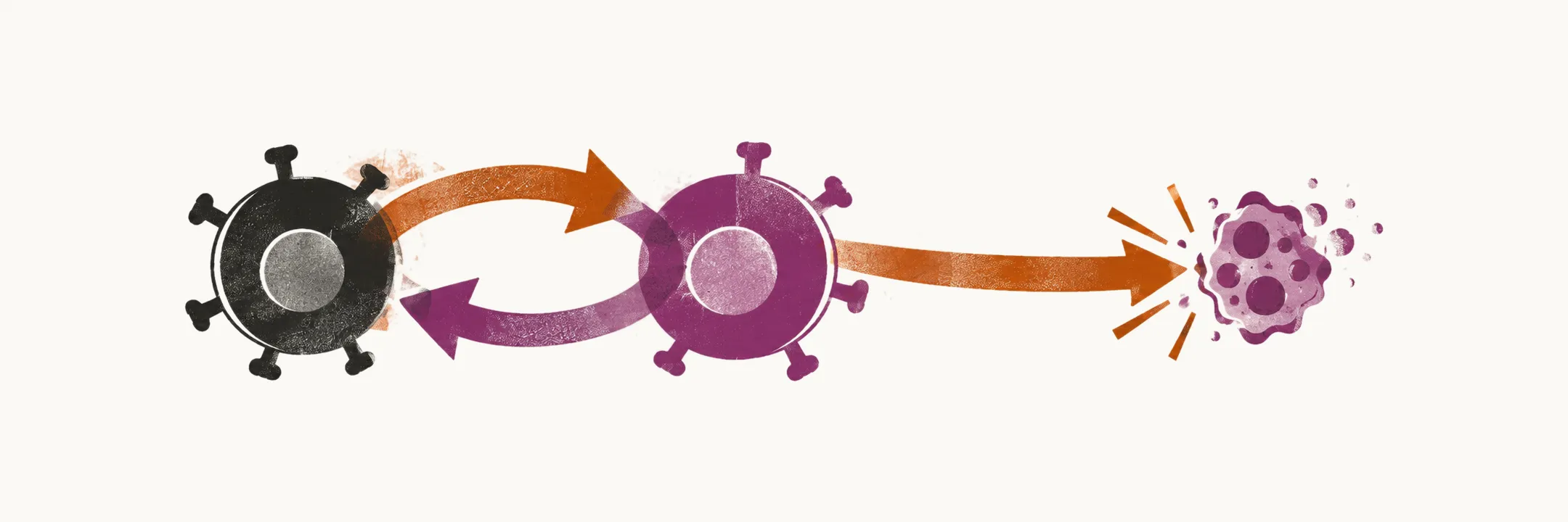
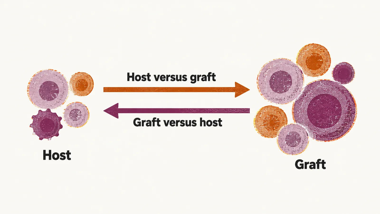
An allogeneic graft, taken from a donor, carries donor immune cells into the patient, and for as long as both sets of cells are present they can recognise each other as foreign. The conflict runs in two directions: host-versus-graft rejection, in which the patient’s immune system attacks the graft, and graft-versus-host disease (GvHD), in which the T cells carried in the graft attack the patient’s tissues. The same donor T cells that cause GvHD can also attack residual malignant cells, and that useful side of the reaction is called the graft-versus-leukemia effect.
Host-versus-graft rejection
The host-versus-graft response is mediated by the residual T cells of the patient, which recognise the HLA (human leukocyte antigen) antigens of the graft as foreign and try to destroy it. In a standard transplant this attack of the host T cells is balanced against the attack of the donor T cells, so neither side acts unchecked. Graft rejection is prevented or managed in two ways: before the transplant, with an immunosuppressive conditioning regimen containing anti-T lymphocyte globulin (ATG) and fludarabine, and after it, with maintained immunosuppression in the recipient, mainly calcineurin inhibitors.
Graft-versus-host disease
GvHD is mediated by the T cells that travel in the graft from the donor into the patient’s body. Donor T cells first recognise the HLA antigens of the recipient’s cells, mainly on dendritic cells (DCs), the cells that present those antigens to the immune system. That recognition activates and expands the donor T cells against the patient’s tissues.
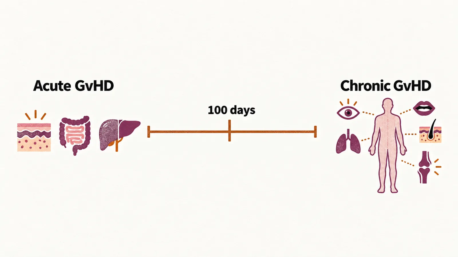
GvHD takes two main forms, separated by when it appears relative to engraftment, the point at which the transplanted stem cells begin to produce blood cells:
- Acute GvHD, appearing less than 100 days after engraftment, affects mainly the skin, the gastrointestinal (GI) tract and the liver, and occurs in 25-30% of allogeneic HSCTs. In the skin it produces a maculopapular rash, in the gut diarrhea, and in the liver a rise in bilirubin and liver enzymes.
- Chronic GvHD, appearing mostly more than 100 days after engraftment (mostly 3-7 months after the transplant), can affect virtually all organs, and occurs in more than 45% of allogeneic HSCTs.
Risk factors for GvHD
How severe GvHD becomes depends first on how closely the donor and recipient HLA types match, then on a sex difference between donor and recipient, and on the source of the HSCs: GvHD happens more with peripheral blood-derived HSCs than with bone marrow or cord blood. Other factors also matter: the conditioning regimen, the age of the recipient and the effectiveness of the GvHD preventive medication, as well as a second transplant or a previous history of GvHD in the patient.
Preventing and treating GvHD
Because GvHD is common and serious, it is prevented rather than simply awaited. Standard prophylaxis is a calcineurin inhibitor combined with a short course of methotrexate, with ATG added when the donor is unrelated. When acute GvHD develops despite prophylaxis, the first-line treatment is systemic corticosteroids.
The graft-versus-leukemia effect
The donor T cells that damage the host can also remove malignant cells that survived the myeloablative procedure. This is what makes GvHD a double-edged sword: a low grade and low stage of damage by the graft to the host, as in stage I GvHD, is associated with more complete removal of the remaining cancer cells, and the survival of patients with grade I GvHD is better than that of patients with grade zero. As the grade of damage rises, this benefit is outweighed by the harm GvHD does to the body.
This phenomenon is called graft-versus-leukemia, or GVL, and it is the useful side of GvHD. A transplant therefore works best when the balance favours GVL rather than severe GvHD.