
In myelodysplastic syndrome (MDS) the blood counts fall while the bone marrow is often hypercellular, and the diagnosis is built from the blood count, the smear and the marrow. The low counts and the busy marrow are the same phenomenon seen from two ends: the marrow is making cells, but they are dysplastic and die before they reach the blood.
Laboratory studies
The blood count, the peripheral smear (blood cells examined under the microscope) and the bone marrow aspirate each contribute to the picture. The findings are described below.
Blood
Anemia is the most common sign, and it is either isolated or in the form of pancytopenia (a low count of red cells, white cells and platelets together). Isolated anemia is common, but isolated neutropenia or thrombocytopenia is not common. Pancytopenia can be found in more than half of the patients at the first diagnosis. Thrombocytopenia is an uncommon complication that can be seen in early MDS, and thrombocytosis (a high platelet count) is very rare, related to del(5q) and to MDS with ring sideroblasts and mutated SF3B1.
The red cells show the features of marrow failure. As in most forms of marrow failure, there is macrocytosis (abnormally large red cells), and the RDW (red-cell distribution width, a measure of variation in cell size) often increases, which reflects anisocytosis (unequal cell size). There is also a low number of reticulocytes, the newly released young red cells.
The other cell lines look abnormal as well. Platelets are large and without granules. Neutrophils are hypogranulated; they can be hypersegmented or even hyposegmented, with the presence of Dohle’s bodies. The count of WBCs is normal or slightly low, except in the case of chronic myelomonocytic leukemia (CMML).
Bone marrow
In 80% of the cases the bone marrow is hypercellular (more cellular than expected for age), but in 20% of the cases it is hypocellular, which leads to confusion with aplastic anemia. The marrow shows a low number of granulocyte precursors and dyserythropoietic changes, which means abnormal development of the red-cell line, and possibly the presence of sideroblastic precursors of RBCs (red-cell precursors with iron deposits ringing the nucleus). Megaloblastic nuclei and defective hemoglobinisation in the erythroid lineage are common.
As a summary, the finding of the bone marrow aspiration would be dyserythropoiesis, dysgranulation and dysmegakaryopoiesis, that is, abnormal development of the red-cell, granulocyte and platelet-producing lines. Mild to moderate myelofibrosis (scarring of the marrow) is reported in more than half of the cases of MDS.
Prognosis is strongly correlated with the proportion of marrow blasts, the immature cells whose percentage also sets the diagnostic boundary below.
Diagnosis
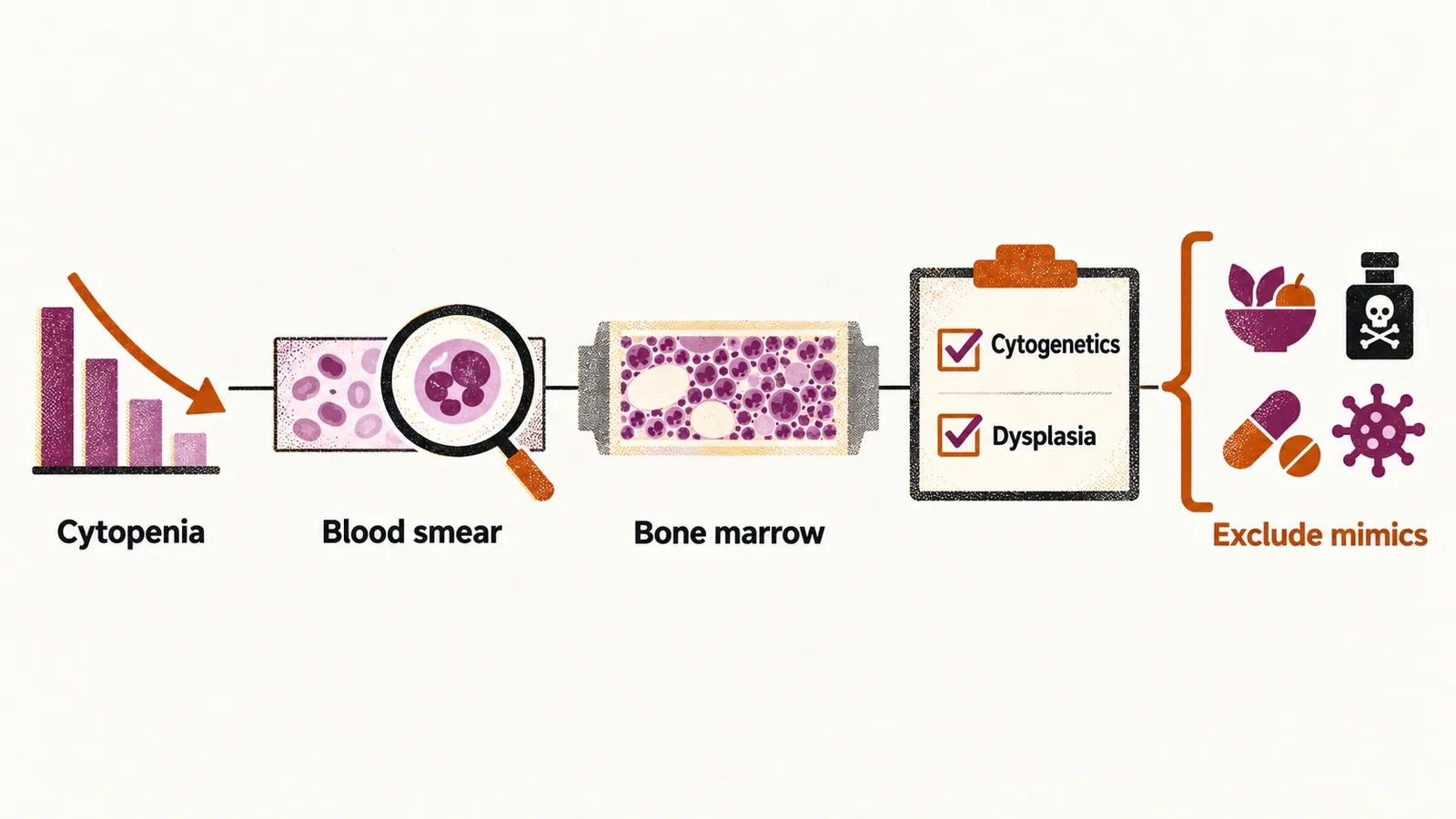
The laboratory findings are turned into a diagnosis by criteria. The diagnosis of MDS should be considered in all patients who have an unexplained persistent cytopenia, and the diagnosis is positive if:
- there is a persistent cytopenia that is not explained by any other pathology, such as infection or vitamin deficiency
- there are fewer than 20% blast cells in the bone marrow
- there are either cytogenetic abnormalities or morphological dysplasia in the blood cells
Differential diagnosis
The differential diagnosis of MDS should be made carefully, in the context of disorders that are cytopenic, dysplastic or clonal problems. Nutritional deficiency, toxic exposure, drugs, infection and a congenital cause can each reproduce part of the picture, and several of them are reversible, which is why they are excluded before the diagnosis is made.
The differential diagnosis in the context of dysplasia and cytopenia would be:
- nutritional deficiencies: vitamin B12 deficiency, folate deficiency, copper deficiency, zinc excess
- toxic exposure: alcohol, heavy metals
- drugs and biologic agents: tacrolimus, valproic acid, mycophenolate mofetil, isoniazid, ganciclovir
- infections: HIV
- congenital disorders
- sideroblastic anemia
Once MDS is established, the question becomes how the patient should be managed, which depends on how advanced the disease is.