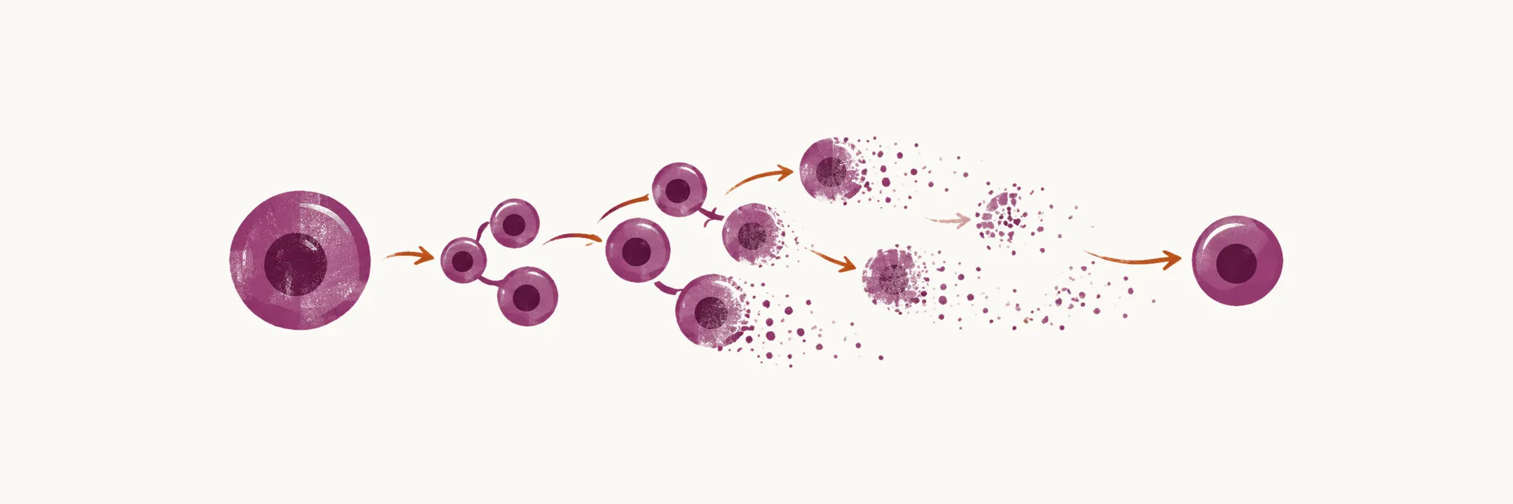
Myelodysplastic syndrome (MDS) is mostly a disease of older adults, and aging is central to its biology. Explaining why a particular patient developed it means separating the few who were exposed to a known cause or carry an inherited syndrome from the majority, in whom the only clear risk factor is time.
Epidemiology
MDS is a disease of the elderly, with onset over 70 years of age. Its incidence in the general population is about 3-20 in 100,000, and the rate is increasing, which is largely due to better diagnosis by physicians rather than to a true change in the disease. MDS is very rare in children, where it is mostly seen in children who carry predisposing genes that can be found by screening for myeloid cancer. In adults, exposure to some chemotherapy agents, such as alkylating agents, and to radiation can also cause it. The exposures are described in the next section.
What causes MDS
The cause is unknown in most cases. When a cause can be identified, MDS is called secondary, and the known contributors fall into two groups: exposures and inherited conditions.
The environmental agents implicated include radiation, benzene, radiotherapy, alkylating agents (busulfan, nitrosourea, procarbazine, anthracyclines and epipodophyllotoxins), and DNA topoisomerase inhibitors. Smoking also raises the risk, roughly doubling it. Separately, some drugs and previous bone marrow disorders such as Fanconi anemia are associated with the disease.
A second group is inherited. Some inherited bone marrow failure syndromes, which include Fanconi anemia and constitutional marrow failure disease, can evolve into MDS.
Despite all of this, most patients with MDS have had no exposure to the environmental risk factors and no previous aplastic anemia (a marrow failure disease with a hypocellular marrow). That is the observation that defines the disease as a disease of aging, consistent with the accumulation of mutations in hematopoietic stem cells (HSCs), the marrow cells from which all blood cells arise.
The mechanism of the disease
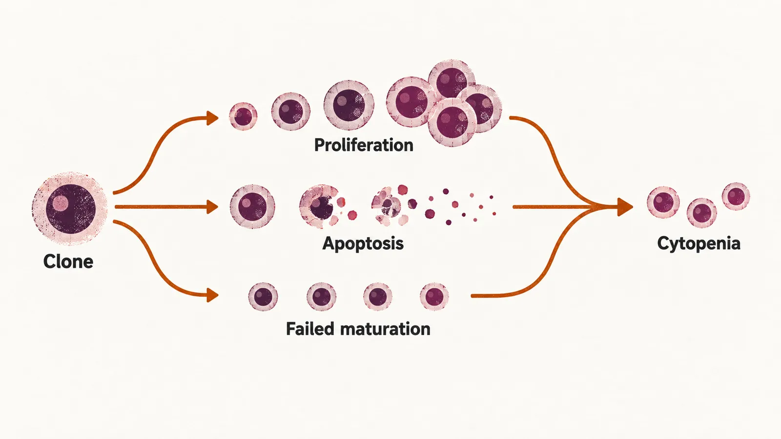
Following the age-related conclusion, the definition of MDS can be refined: it is a clonal hematopoietic stem cell disorder characterised by disordered proliferation, differentiation and aberrant hematopoiesis, which leads to cytopenia and a risk of progression to leukemia. Clonal means that the abnormal cells descend from one mutated stem cell.
The paradox that a low blood count coexists with a busy marrow is explained by three cardinal features that can be used for diagnosis:
- increased marrow proliferation (cells multiply)
- increased marrow apoptosis (programmed cell death)
- decreased marrow differentiation (cells fail to mature into functioning blood cells)
The clone expands and fills the marrow, but its cells die before they mature and leave the marrow, so the peripheral counts fall. Cytogenetic abnormalities, meaning changes in the number or structure of chromosomes, also accumulate with age and are part of this picture.
Cytogenetics
Cytogenetic abnormalities are not random, and some follow a specific cause. Aneuploidy (a gain or loss of whole chromosomes) is more common than translocation (a piece of one chromosome joined to another) in MDS, which separates it from many leukemias, where translocations dominate. Loss of chromosome 5 or 7 has been related to previous treatment with alkylating agents, rearrangements of 11q23 follow topoisomerase II inhibitors, and loss of chromosome 20 and trisomy of 8 are also seen in MDS.
The type, and also the number, of cytogenetic abnormalities in MDS is strongly correlated with the chance of leukemic transformation of MDS. This is why karyotype contributes as much to prognosis as the blast count.
Recurrent mutations and their prognostic weight
Karyotype describes whole chromosomes; mutations describe changes within individual genes. Some of the mutations found in MDS are also found in acute myeloid leukemia (AML), but some are unique to MDS. A few carry enough prognostic weight to be remembered by name. The SF3B1 mutation is associated with a good prognosis, whereas mutations of RUNX1, TP53, EZH2 and ASXL1 are associated with a poor one.
Inherited (germline) changes matter too. A family history of sideroblastic anemia, Fanconi anemia, telomeropathy, or an inherited GATA2 mutation in MonoMac syndrome suggests that a patient’s cytopenia may be progressing to MDS. A RUNX1 germline mutation also confers a high risk of MDS and leukemia, and it is preceded by years of modest thrombocytopenia.
When a genetic finding is enough for a diagnosis
Cytogenetic and molecular findings can also settle the diagnosis. Some, when accompanied by cytopenia, are enough to indicate a diagnosis of MDS:
- a mutation in the SF3B1 gene
- a 5q deletion with up to one additional cytogenetic abnormality, except the 7q deletion
Other abnormalities mean nothing on their own: del(20q), del(Y) and trisomy 8 do not indicate MDS in the absence of dysplasia or cytopenia, so they cannot be used to make the diagnosis by themselves.
Finally, some abnormalities exclude MDS because they are diagnostic of AML:
- t(8;21)
- t(15;17)
- inv(16)
These findings describe the clone itself. What the patient experiences comes from the cytopenias the clone produces, which is the question of clinical presentation.