Hypoventilation means breathing that is too shallow or too slow to clear carbon dioxide. It is the problem that ventilatory support is designed to treat, and it is the reason oxygen alone is not the answer: oxygen corrects the saturation reading without correcting the carbon dioxide that accumulates underneath.
Why oxygen alone can be dangerous
A neurological patient with a weak cough retains secretions and arrives with low oxygen saturation. That low saturation reflects hypoventilation from muscle weakness rather than primary lung disease, and the remaining ventilatory drive may depend substantially on the hypoxic stimulus — the low oxygen itself is part of what keeps the patient breathing. Giving supplemental oxygen alone can blunt that drive and allow progression toward hypercapnic respiratory failure, in which carbon dioxide accumulates. The correct response is ventilatory support combined with secretion clearance under local protocols, not oxygen alone.
The same hazard is explained in the swallowing context in Neurological Dysphagia — Respiratory Planning and Nutrition.
Starting non-invasive ventilation
Non-invasive ventilation (NIV) supports breathing through an interface such as a mask or mouthpiece rather than a tube placed in the airway. It relieves the symptoms of respiratory impairment and can prolong life, but it does not stop progression of the underlying disease, which is a distinction that matters when the treatment is discussed with a patient.
Starting it is a structured process: assess indications and contraindications, discuss the proposal with the patient, obtain consent, choose the device and interface, begin with low-pressure adaptation, verify tolerance, and then check effectiveness.
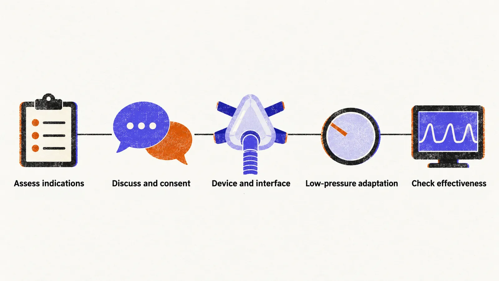
Example adaptation pressures are inspiratory positive airway pressure (IPAP) 8-10 with expiratory positive airway pressure (EPAP) 4-5 cmH2O, used at least 3-4 hours per night while tolerance is verified. These are adaptation examples, not a universal protocol: indications, carbon dioxide thresholds and contraindications differ by disease and guideline, and motor neuron disease guidance sets its own assessment criteria for starting ventilation.
Checking that ventilation is working
Once ventilation has been started, its effectiveness is checked with nocturnal polygraphy or oximetry, waking arterial blood gas, and non-invasive carbon dioxide monitoring. Symptom review for hypoventilation, compliance data (how much the patient actually uses the device), and ventilator download data (the device’s own recorded settings and use) complete the check.
Starting ventilation is a decision made with the patient and reviewed over time, and it works best when the rest of the respiratory plan is already in place, with assessment, secretion clearance and support arranged together.