
Celiac disease is a permanent, immune-mediated intolerance of gluten that damages the mucosa of the small intestine, and its diagnosis starts with serology: blood tests for the antibodies the disease produces. Since 2020 a defined group of children can be diagnosed without a duodenal biopsy at all. Two rules govern the whole pathway. The child must be eating gluten in normal amounts when the tests are taken, because a diet that has already been restricted can normalize both the antibodies and the histology (the microscopic findings on biopsy) and produce a false negative. And the antibody titre — the measured level, not merely its positivity — decides whether the biopsy can be avoided.
Who to test
Testing is indicated for three groups of children:
- Children with suggestive symptoms — chronic diarrhea, faltering growth, unexplained iron-deficiency anemia, and the other presentations described in Celiac Disease in Children.
- Children with associated conditions — type 1 diabetes, autoimmune thyroid disease, Down syndrome, Turner syndrome, Williams-Beuren syndrome, IgA deficiency.
- First-degree relatives of a person with celiac disease.
The last two groups include children with no symptoms at all, which is how the silent form of the disease is found.
Serology
The first-line combination is total IgA plus anti-tTG IgA, the IgA-class antibody against tissue transglutaminase, the enzyme at the centre of the disease mechanism. Total IgA is included because selective IgA deficiency produces false-negative IgA-class results; if total IgA is low, the screening test switches to an IgG-based test (anti-tTG IgG, anti-deamidated gliadin peptide IgG, or EMA IgG). Anti-endomysial antibodies (EMA) are a highly specific confirmatory test used second, and are best performed in an experienced laboratory.
Diagnosis without biopsy
The 2020 guideline of ESPGHAN (the European Society for Paediatric Gastroenterology, Hepatology and Nutrition) allows a diagnosis without duodenal biopsy when anti-tTG IgA is at least 10 times the upper limit of normal and EMA is positive in a second serum sample. The upper limit of normal is the top of the laboratory’s reference range for the assay. Two details matter:
- The threshold is 10 times the upper limit of normal, not simply “positive”. Below that value the false-positive rate is high enough that biopsy is required, even with a positive EMA.
- HLA typing and the presence of symptoms are no longer requirements. The pathway can be used in asymptomatic children, using the same criteria.
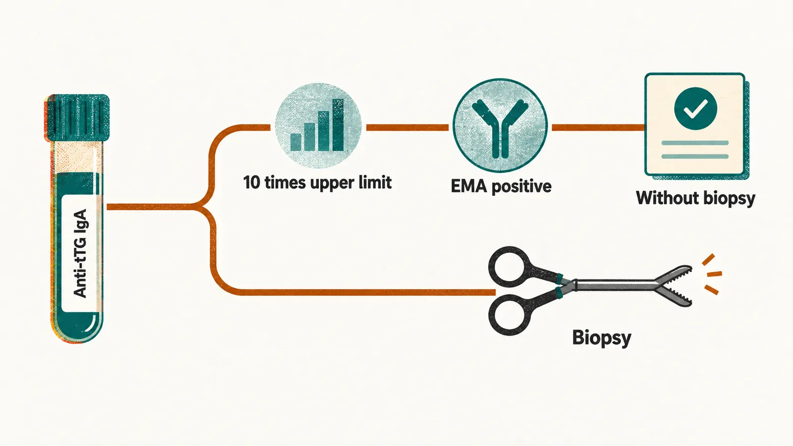
Some laboratories can only report a value above the measuring range; the guideline requires an assay with a calibration curve whose range includes the 10× upper-limit value, because the numeric threshold is what makes the diagnosis.
When biopsy is needed
Biopsy is required whenever serology cannot carry the diagnosis on its own:
- anti-tTG IgA is positive but below 10 times the upper limit of normal;
- the serology is equivocal or discordant;
- EMA is negative;
- the child is IgA-deficient and has tested positive on an IgG-based test, in every case.
Biopsies are taken during a gluten-containing diet, with at least four specimens from the distal duodenum and at least one from the duodenal bulb, because the lesion is patchy and can spare the bulb or spare the distal duodenum.
Clinical judgement matters here as well: when histological proof is likely to be needed later to persuade a family to commit to a lifelong diet, biopsy is preferred even if the serology would permit skipping it.
HLA typing belongs with the equivocal cases above rather than with first-line testing. Because HLA-DQ2 and HLA-DQ8 are absent in only a small minority of people with celiac disease, their absence makes the diagnosis very unlikely, and that is the one question typing answers; the haplotypes themselves are too common in the general population to confirm anything (see Celiac Disease in Children).
Histology
The biopsy is graded with the modified Marsh-Oberhuber classification, from normal mucosa to total villous atrophy, in which the villi (the finger-like projections that absorb nutrients) are completely flattened. Two further terms recur in it: intraepithelial lymphocytes are lymphocytes lying within the surface lining, and crypt hyperplasia means lengthened crypts, the glands between the villi.
| Grade | Findings |
|---|---|
| 0 | Normal mucosa |
| 1 (infiltrative) | Normal villous architecture with increased intraepithelial lymphocytes |
| 2 (hyperplastic) | Increased intraepithelial lymphocytes with crypt hyperplasia, villi still present |
| 3 (destructive) | Villous atrophy with crypt hyperplasia, the typical celiac lesion — subdivided into 3a (mild), 3b (moderate) and 3c (total atrophy) |
A normal endoscopy does not exclude the diagnosis. The bulb and the distal duodenum can look unremarkable while the histology is diagnostic, which is why the biopsy protocol above is specified rather than left to the endoscopist’s impression. Whichever route confirms it, a diagnosis of celiac disease commits the child to a lifelong gluten-free diet.