
Celiac disease is a permanent, immune-mediated intolerance of dietary gluten, the protein found in wheat, barley and rye, in genetically predisposed people, which produces damage to the mucosa (the lining) of the small intestine. Permanent is the word that carries the clinical weight: the mucosa recovers when gluten is removed, but the intolerance does not go away, which is why the treatment is lifelong rather than a course. A child who is told they have outgrown celiac disease has either been misdiagnosed or has abandoned the diet.
Celiac disease is the malabsorptive arm of chronic diarrhea in a child: the child who presents with prolonged loose stools, faltering growth and nutritional deficiencies, and whose first-line tests point to the duodenum rather than the colon.
How common it is
Celiac disease is common enough that it belongs in the differential of almost any child with chronic diarrhea, short stature or unexplained iron-deficiency anemia. In a pooled analysis of 275,818 people screened by serology (blood tests for the antibodies of the disease), the global prevalence was 1.4% (95% confidence interval, CI, 1.1–1.7%), and biopsy-confirmed disease was present in 0.7% (95% CI 0.5–0.9%) of 138,792 people tested. Prevalence was higher in children than in adults in the same analysis.
Most people with celiac disease are undiagnosed, because the classic diarrheal presentation is now the minority. Italy has taken a different route from most countries: a 2023 national law provides for population screening of children for celiac disease and type 1 diabetes. The school-age study behind that policy found an overall prevalence of 1.65% (95% CI 1.34–2.01%), with only about 40% of the affected children already diagnosed before screening. Other European countries have not adopted universal screening, so the Italian programme is a policy choice rather than a settled international standard.
Screening is a different question from prevention, and no feeding strategy prevents celiac disease. Introducing gluten at any point between 4 and 12 months of age does not change the overall risk, breastfeeding does not lower it, and no gluten dose has been shown to prevent the disease, so infant feeding is not modified with prevention in mind — including in a child with an affected first-degree relative.
The mechanism in four steps
The disease arises where gluten meets two things: an enzyme called tissue transglutaminase (tTG), and the HLA (human leukocyte antigen) molecules that display peptide fragments to T cells.
- Gluten is partially digested in the lumen into gliadin peptides that resist further breakdown.
- Tissue transglutaminase deamidates glutamine residues on those peptides, converting them to glutamic acid.
- The deamidated peptides bind with high affinity to HLA-DQ2 or HLA-DQ8 on antigen-presenting cells and are presented to CD4+ T lymphocytes (helper T cells).
- T-cell activation, with IL-15 (interleukin-15) signalling from the epithelium, drives a Th1-type inflammatory response that flattens the villi (the finger-like projections that absorb nutrients), lengthens the crypts (the glands between them) and destroys enterocytes (the absorptive cells).
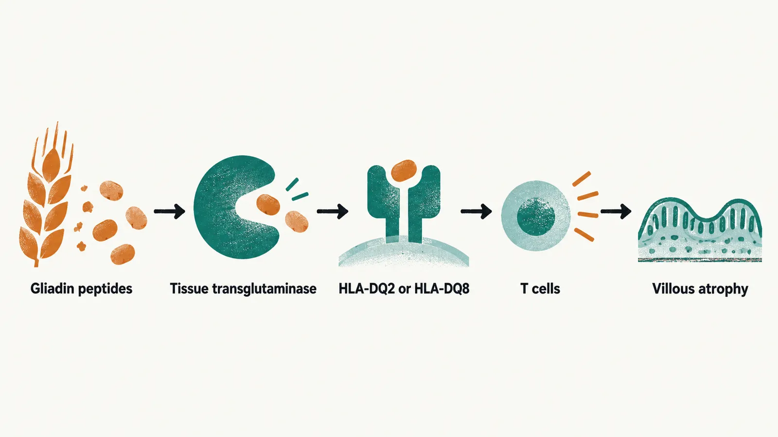
The same enzyme that modifies the peptide is the target of the autoantibody response, which is why anti-tTG IgA, the IgA-class antibody against tissue transglutaminase, is the serological marker of the disease. Serology tracks the autoimmune process, not gluten intake directly, and falls as the mucosa heals on treatment.
Genetics — a rule-out test, not a diagnosis
Step 3 depends on the class II HLA haplotypes HLA-DQ2 and HLA-DQ8, inherited combinations of HLA genes with which celiac disease is strongly associated. An important asymmetry follows from how common those haplotypes are:
- HLA-DQ2 or HLA-DQ8 is present in roughly 98% of people with celiac disease (in European patients, DQ2 in about 90% and DQ8 in about 5–10%).
- The same haplotypes are carried by a large minority of healthy people. Reported frequencies in general populations range from roughly 30% to over 50%, depending on the population studied.
- Absence of both haplotypes makes celiac disease very unlikely.
HLA typing therefore has one main use — excluding the diagnosis in a child whose serology is equivocal or whose biopsies were uninformative. A positive HLA result means the child belongs to a large group most of whom never develop the disease, and never confirms it. The 2020 ESPGHAN guideline reflects this by no longer requiring HLA typing for a serology-based diagnosis.
Clinical forms
The disease is recognized in four forms, and only the first is defined by intestinal symptoms. The others explain why so many affected children are found through growth, blood tests or screening rather than through diarrhea.
| Form | What it looks like |
|---|---|
| Typical (classic) | Diarrhea, weight loss, anorexia, abdominal pain and distension, vomiting, sometimes irritability or personality change. Onset follows gluten introduction. Once the usual pediatric presentation; now the minority |
| Atypical (non-gastrointestinal) | Consequences of malnutrition dominate — short stature, iron-deficiency anemia that does not respond to oral iron, osteopenia, dental enamel defects. Intestinal symptoms may be absent or minimal |
| Silent | Villous atrophy (flattened villi) with positive serology in a child who has no symptoms at all; found through screening of relatives, children with type 1 diabetes, or a population programme |
| Potential | Positive serology with normal or minimally abnormal histology (the microscopic appearance of the biopsy). This is a pre-stage that may evolve, so it needs surveillance rather than dismissal |
Growth failure is the feature that most often gives the diagnosis away in a child. Celiac disease should be suspected in any infant or child who is not growing as expected, and in any child with iron-deficiency anemia that fails to respond to iron replacement. Recurrent aphthous stomatitis, abdominal pain and diarrhea together raise the possibility of both celiac disease and IBD, which is why the two pathways overlap in the work-up — the inflammatory branch is set out in Pediatric Inflammatory Bowel Disease.
Dermatitis herpetiformis is a separate presentation of the same disease: an intensely itchy, chronic, relapsing rash of small papules and vesicles in clusters on the extensor surfaces, buttocks, shoulders, trunk, face and scalp. It may occur with no intestinal symptoms at all. When it is present, it confirms the diagnosis, and the diagnosis should be confirmed histologically by skin biopsy rather than assumed, because other itchy vesicular eruptions occur in childhood.
The diagnosis starts with serology, and ends with histology in the children who need a biopsy; both pathways — with and without a duodenal biopsy — are set out in Diagnosing Celiac Disease in Children, and the diet and follow-up that come after a diagnosis are in Treating and Monitoring Celiac Disease.
Celiac disease and its neighbours
Three conditions share gluten as a trigger and are frequently confused.
| Disorder | Mechanism | How it is identified |
|---|---|---|
| Celiac disease | Autoimmune Th1 enteropathy with villous atrophy | Anti-tTG IgA and EMA (anti-endomysial antibodies), with or without biopsy |
| Wheat allergy | IgE-mediated (type I) hypersensitivity | Skin prick test or specific IgE; symptoms on exposure, including after inhalation |
| Non-celiac wheat sensitivity | No identified autoimmune or allergic mechanism | Clinical; no diagnostic test, no mucosal damage, no autoantibodies |
The distinction matters because the treatments differ: celiac disease requires gluten exclusion for life; wheat allergy requires wheat avoidance with attention to anaphylaxis risk; and non-celiac wheat sensitivity is managed by clinical improvement on a restricted diet, with the caveat that expectation effects make it difficult to assess objectively. The evidence for the gluten-related disorder spectrum is set out in the European Society for the Study of Coeliac Disease guideline.