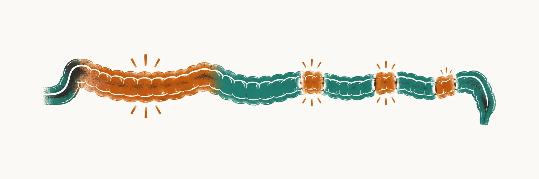
Inflammatory bowel disease (IBD) is chronic, relapsing inflammation of the gastrointestinal tract of unknown cause, classified by its anatomical and histological pattern into ulcerative colitis (UC), Crohn’s disease (CD) and IBD-unclassified (IBDU). In children it behaves differently from the adult disease in two ways that shape everything else: the presentation is often subtle and slowly progressive rather than dramatic, and the inflammation interferes with growth during the years when growth is the point. Both push the diagnostic threshold down, so the working rule in pediatric IBD is that the more carefully you look, the more you find.
IBD is the inflammatory arm of chronic diarrhea in children; the malabsorptive arm is Celiac Disease in Children.
Ulcerative colitis, Crohn’s disease and the unclassified middle
The two main forms differ in where the inflammation sits and how deep it goes.
| Feature | Ulcerative colitis | Crohn’s disease |
|---|---|---|
| Distribution | Continuous, starting at the rectum and extending proximally | Patchy, with skip lesions (normal bowel between affected segments), anywhere from mouth to anus |
| Depth | Mucosa only | Transmural, through the full thickness of the bowel wall |
| Typical site | Colon | Terminal ileum, colon, upper gastrointestinal tract, perianal region |
| Ulcers | Superficial | Deep and linear |
| Complications | Acute severe colitis, eventually colectomy | Strictures (narrowed segments), fistulas (abnormal tracts between bowel and other organs or skin), abscesses, perianal disease |
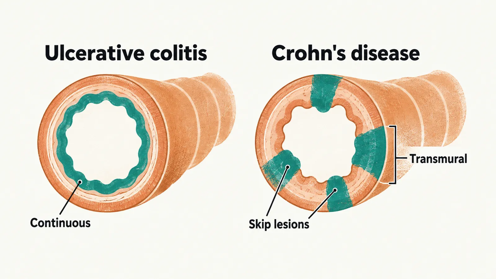
IBDU describes disease with features of both at diagnosis, and it is a real category rather than a diagnostic failure: some of these children are reclassified as UC or CD as the disease declares itself over time. The Paris classification, a pediatric modification of the Montreal classification, is the current pediatric standard for describing phenotype, meaning where the disease sits and how it behaves.
The practical consequence of the table is that perianal disease — skin tags, fissures, fistulas, abscesses — points to Crohn’s disease, because ulcerative colitis does not cause it. Upper gastrointestinal involvement is also a Crohn’s pattern, and it is common enough in children that the diagnostic endoscopy includes the upper tract even when the symptoms are colonic.
Who gets it, and when
Pediatric-onset IBD is being diagnosed more often, and the increase is largest in the youngest children. Very early onset IBD, defined as onset before 6 years of age, accounts for roughly 3–15% of pediatric IBD across reported cohorts. Onset before 2 years is a distinct problem: a substantial minority of these children have a monogenic defect (a disorder caused by a single gene) of immunity or epithelial function rather than idiopathic IBD, with more than 70 genes implicated. That is why very early onset disease — particularly with atypical features such as severe perianal disease, recurrent infection or a family history suggesting immunodeficiency — prompts genetic testing and specialist referral rather than a standard work-up.
When to suspect IBD in a child
Because the pediatric presentation is often subtle and slowly progressive, the trigger set is broader than bloody diarrhea:
- Chronic diarrhea with blood or mucus, with abdominal pain, urgency and tenesmus (a painful, persistent urge to pass stool).
- Unexplained delay in weight gain or in linear growth, especially with fever or laboratory markers of malnutrition or inflammation.
- Chronic diarrhea without blood or mucus, particularly if it occurs at night and is accompanied by systemic signs or abnormal laboratory results.
- Severe, disabling abdominal pain with nocturnal symptoms and malnutrition.
- Any of the above together with extraintestinal manifestations — arthritis, uveitis, erythema nodosum, pyoderma gangrenosum, chronic liver disease.
Growth failure and nocturnal diarrhea are the two items that carry the most weight in children, because both are unusual in the functional and benign patterns. Which symptoms appear also depends on where along the gut the disease sits.
| Location | Dominant presentation |
|---|---|
| Stomach and duodenum | Nausea, vomiting, epigastric pain, poor appetite |
| Small bowel | Malabsorption with chronic diarrhea and weight loss, pain, fever; sub-occlusion with vomiting and distension |
| Colon | Bloody diarrhea, urgency, tenesmus, pain |
| Perianal | Skin tags, fissures, fistulas, abscesses |
Extraintestinal manifestations
IBD is not confined to the intestine. The extraintestinal features listed among the triggers above sometimes precede the gastrointestinal ones and bring the child to medical attention first, so it helps to know them by organ.
- Skin — erythema nodosum (tender red nodules under the skin) and pyoderma gangrenosum (painful ulcerating skin lesions).
- Joints — arthritis, arthralgia, sacroiliitis (inflammation of the joints between the spine and pelvis).
- Eyes — uveitis, episcleritis, iritis.
- Liver and biliary tract — including primary sclerosing cholangitis, a chronic inflammatory disease of the bile ducts.
- Mouth — recurrent aphthous ulcers.
- Kidney — nephrolithiasis (kidney stones).
Erythema nodosum and pyoderma gangrenosum are associated with IBD but not specific to it; erythema nodosum also occurs in streptococcal infection, sarcoidosis and tuberculosis, and pyoderma gangrenosum in other immune-mediated diseases.
Diagnosis
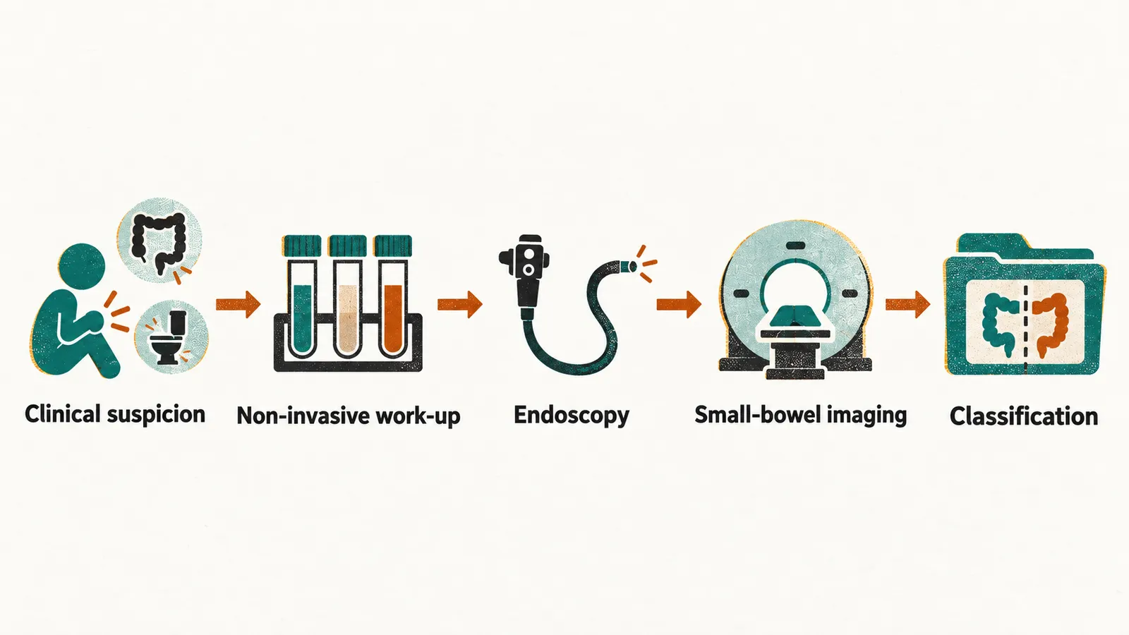
The European framework is the revised Porto criteria of ESPGHAN (the European Society for Paediatric Gastroenterology, Hepatology and Nutrition), which require the whole gastrointestinal tract to be assessed before IBD can be classified. In practice the pathway runs as follows.
- Clinical suspicion from the trigger set above.
- Non-invasive work-up — blood count, CRP and ESR, iron studies, albumin and nutritional markers, and fecal calprotectin (explained below), with abdominal ultrasound.
- Endoscopy — upper gastrointestinal endoscopy and ileocolonoscopy (colonoscopy that continues into the terminal ileum), with segmental biopsies from the terminal ileum and every colonic segment.
- Small-bowel imaging — magnetic resonance enterography (MRI of the small bowel), ultrasound or capsule endoscopy (a swallowed camera capsule) when small-bowel involvement is suspected, and in all children unless the endoscopic and histological picture is typical of ulcerative colitis.
- Classification as UC, CD or IBDU from the combined endoscopic, histological and imaging findings.
Skipping the upper endoscopy in a child with suspected IBD is a known way to miss upper gastrointestinal Crohn’s disease, and skipping small-bowel imaging is a known way to miss small-bowel disease that the colonoscope cannot reach.
Fecal calprotectin
Fecal calprotectin is a calcium- and zinc-binding protein that makes up a large share of neutrophil cytosolic protein. When intestinal mucosa is inflamed, neutrophils (the white cells of acute inflammation) migrate into the lumen and release it, so the stool concentration reflects the intensity of neutrophilic inflammation. It is the most useful non-invasive biomarker in pediatric IBD, and it is also unspecific.
Reference values fall steeply with age. In the first year of life, values of several hundred µg/g can be found in healthy infants; from about 4 years of age the upper limit of normal is usually taken as roughly 50 µg/g. Published age-banded upper limits vary substantially between studies — one healthy-child dataset gives 910 µg/g at 0–12 months, 286 µg/g at 1–4 years and 54 µg/g at 4–12 years, while other cohorts report much lower figures — so a result has to be read against the age band and the clinical picture rather than against a single cutoff.
Raised values also occur in bacterial and viral gastroenteritis, Giardia infection, food allergy, cystic fibrosis, untreated celiac disease, use of NSAIDs or proton-pump inhibitors, and in young children generally. Untreated celiac disease raises fecal calprotectin, which is the practical reason a raised value in a child with chronic diarrhea does not establish IBD until celiac serology has been checked. The first bowel movement of the day is the preferred sample, and assay choice and sample handling both affect the result.
Prognosis at diagnosis
Classification says what the disease is; the findings at diagnosis also suggest how it will behave. Several features at diagnosis predict a more aggressive course in pediatric Crohn’s disease: extensive ileal involvement, perianal disease, stricturing behavior (disease that narrows the bowel), deep colonic ulcers, upper gastrointestinal involvement, a high corticosteroid requirement at presentation, and high serological titers. These are the features that push treatment decisions toward earlier and more effective therapy rather than a slow step-up, and they are the reason the extent of disease is mapped at diagnosis rather than assumed. The treatment this mapping leads to is set out in Treatment and Monitoring of Pediatric Inflammatory Bowel Disease.