
Chronic diarrhea in a child means loose stools that continue for 14 days or more. Its causes run from benign functional patterns to celiac disease, inflammatory bowel disease (IBD) and congenital defects, so the work-up has to find the children with organic disease without investigating every child. Throughout, the aim is to decide which mechanism is producing the stools — osmotic, secretory, fat malabsorption or inflammation — because the mechanism decides which part of the work-up to follow.
The work-up is therefore a sequence rather than a panel: history and growth chart first, then a targeted examination, then first-line non-invasive tests, and endoscopy only when those tests point somewhere. The purpose of the gating is that most children do not need an endoscopy, and the ones who do should have the right one — a duodenal biopsy and a colonic biopsy answer different questions.

Step 1 — History
The history decides the pre-test probability — how likely each diagnosis is before any test is done — of almost everything that follows. It starts with the stools themselves and widens outwards to the child’s diet, exposures and family.
- Onset and pattern. When did the loose stools start, and did they begin after an acute gastroenteritis that never fully resolved? Is the pattern continuous or intermittent? A duration of 14 days or more makes the episode chronic.
- Stool characteristics. Consistency, frequency, color, smell, presence of undigested food, blood or mucus, and the relationship to meals. These are the observations that map onto the mechanisms in Definitions and Mechanisms of Chronic Diarrhea in Children: greasy and pale suggests fat malabsorption, bloody and mucousy suggests inflammation, acidic stools after dairy suggest carbohydrate malabsorption.
- Associated symptoms. Abdominal pain, vomiting, fever, rash, joint symptoms, mouth ulcers, perianal problems, weight loss and fatigue.
- Diet. Age at gluten introduction and the amount of gluten eaten, the type and volume of milk, fruit juice intake, recent formula changes, and any food the family has already removed. A diet that has been restricted before assessment can normalize serology (the blood antibody tests) and histology (the biopsy findings), so it matters what the child was eating when the symptoms started.
- Drugs and exposures. Antibiotics, laxatives, recent travel, day-care attendance.
- Family history. First-degree relatives with celiac disease, type 1 diabetes, autoimmune thyroid disease or IBD all raise the probability of the corresponding diagnosis in the child.
Step 2 — Growth chart and examination
The growth chart is the single most informative non-invasive assessment, and it is the part that separates a benign functional pattern from organic disease.
- Growth below the 2nd centile for age is abnormal, and a fall across two or more weight centile spaces (the bands between adjacent centile lines on the chart) is a red flag even when the absolute centile still looks acceptable. For a child whose birth weight was above the 91st centile the corresponding threshold is three centile spaces.
- Weight deceleration out of proportion to height suggests recent-onset organic disease, because weight falls before length does.
- Serial measurements matter more than a single point: a chart with two or three points in it can show a trajectory, and one point cannot.
Once the growth pattern is known, the examination runs from head to toe, looking for the systemic signs of malabsorption, anemia, inflammation and dehydration.
- General. How unwell the child looks; pallor from anemia; edema from hypoalbuminemia (low blood albumin), which occurs in protein-losing states including celiac disease.
- Skin. Dermatitis herpetiformis, the intensely itchy papulovesicular rash of celiac disease on the extensor surfaces, and the IBD-associated erythema nodosum (tender red nodules under the skin) and pyoderma gangrenosum (painful ulcerating skin lesions).
- Eyes and mouth. Uveitis and episcleritis (inflammation of the inner layers and of the outer coat of the eye) and recurrent aphthous stomatitis (mouth ulcers), which occur in both celiac disease and IBD.
- Abdomen. Distension, tenderness, organomegaly, masses, and the hard fecal mass of overflow around constipation.
- Perianal inspection. Skin tags, fissures, fistulas (abnormal tracts opening onto the skin) and abscesses, which point to Crohn’s disease.
- Growth and pubertal staging, documented rather than estimated.
Red flags
History, growth chart and examination together answer the first question of the work-up: is this a reassuring pattern, or does the child need investigating? Any one of these features turns a reassuring pattern into an indication to investigate:
- Weight loss or growth faltering — the most important single sign.
- Blood or mucus in the stool.
- Nocturnal diarrhea, particularly in a child who was previously continent. Diarrhea that wakes a child from sleep suggests organic disease rather than a functional pattern.
- Fever, rash, arthritis or recurrent vomiting — systemic features.
- Abnormal laboratory results: raised CRP or ESR, anemia, or signs of chronic inflammation.
- A family history of IBD or celiac disease combined with any of the above.
Step 3 — First-line non-invasive tests
The first-line tests are read as a pattern rather than one at a time. Blood tests look for anemia, protein loss, inflammation and vitamin deficiency; celiac serology screens for celiac disease; and stool tests look for inflammation, pancreatic insufficiency, carbohydrate malabsorption, protein loss and infection.
| Test | What it is for |
|---|---|
| Complete blood count, ferritin, iron studies, folate, vitamin B12 | Anemia and its pattern: microcytic iron-deficiency anemia suggests blood loss or malabsorption; macrocytic anemia suggests B12 or folate malabsorption |
| Albumin and total protein | Protein-losing enteropathy or severe malabsorption; a low albumin with edema is an organic marker |
| CRP and ESR (C-reactive protein and erythrocyte sedimentation rate, blood markers of inflammation) | Inflammation; a normal CRP with a low albumin and low iron argues for malabsorption rather than active inflammation |
| Vitamin D, and calcium if abnormal | Fat-soluble vitamin status in fat malabsorption |
| Total IgA and anti-transglutaminase IgA | First-line celiac screening; anti-transglutaminase IgA is the celiac antibody, and total IgA is needed because selective IgA deficiency produces false-negative IgA results (see Celiac Disease in Children) |
| Stool occult blood | Mucosal inflammation or bleeding |
| Fecal calprotectin | Neutrophil-derived marker of intestinal inflammation; interpretation is age-dependent and it rises in several conditions other than IBD (see Pediatric Inflammatory Bowel Disease) |
| Fecal elastase-1 | A pancreatic enzyme measured in stool; tests for pancreatic exocrine insufficiency when stools are pale and greasy |
| Stool pH and reducing substances | Carbohydrate malabsorption; an acidic stool with reducing substances is the pattern |
| Stool alpha-1 antitrypsin | A blood protein that appears in stool when protein leaks through the gut wall; tests for protein-losing enteropathy, when albumin is low and there is no other explanation |
| Parasitology, and stool culture where indicated | Persistent giardiasis (Giardia infection) and other infections that mimic malabsorption |
When fat malabsorption is suspected, the first-line panel is completed by a sweat chloride test for cystic fibrosis, the most important cause of pancreatic exocrine insufficiency; a value of 60 mmol/L or more confirms cystic fibrosis and a value below 30 mmol/L makes it unlikely.
The pattern these tests produce is what the next step depends on. A typical malabsorptive picture is a microcytic anemia with low ferritin, low albumin, low fat-soluble vitamin levels and a normal CRP. A typical inflammatory picture is a raised CRP or ESR, raised fecal calprotectin and blood in the stool.
Step 4 — Targeted endoscopy
Endoscopy is gated by the non-invasive results, and the mechanism pattern chooses the route.
- Malabsorptive pattern — low albumin, iron or vitamin deficiencies, steatorrhea, normal inflammatory markers, no blood in the stool: upper gastrointestinal endoscopy with duodenal biopsies, which is the route to celiac disease and other mucosal causes.
- Inflammatory pattern — blood or mucus in the stool, raised CRP or fecal calprotectin: ileocolonoscopy with biopsies from the terminal ileum and every colonic segment, which is the route to IBD.
- Unclear or mixed pattern: both procedures, with small-bowel imaging by ultrasound, magnetic resonance enterography (MRI of the small bowel) or capsule endoscopy (a swallowed camera capsule) when small-bowel Crohn’s disease is a possibility.
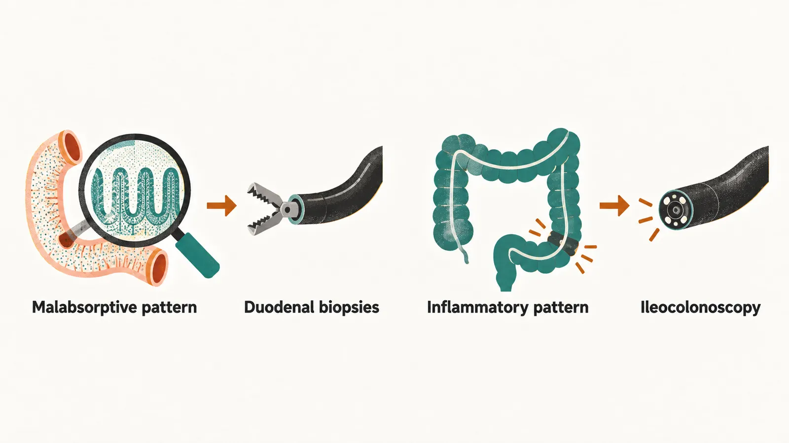
A normal-looking duodenum does not exclude celiac disease, because the lesion is patchy and the diagnosis is histological. And an upper endoscopy is part of the pediatric IBD work-up even when the symptoms are colon-predominant, because upper gastrointestinal Crohn’s disease is easy to miss.
Diet and drugs while the work-up is running
Tests and endoscopy take time, and two management questions arise while they run: what the child should eat, and whether a drug should be given to slow the stools.
There is no specific diet that treats chronic diarrhea in children, and restricting the diet before the diagnosis is made can hide the diagnosis. The child stays on an age-appropriate diet. Excessive fruit juice and large volumes of milk can amplify stool output and are worth moderating, and a short trial of a lactose-free formula is reasonable in selected children with severe symptoms. The evidence for that trial, and its limits, is in Refeeding and Drug Therapy in Acute Diarrhea.
Antimotility drugs, which slow intestinal transit, have no place in the management of chronic diarrhea in children. The European ESPGHAN/ESPID acute gastroenteritis guideline states that loperamide is not recommended in children with gastroenteritis, and the concern applies at least as strongly to a chronic picture, where slowing transit masks inflammation, delays diagnosis and risks ileus, a functional obstruction of the bowel. In functional diarrhea in particular there is no toxic hypersecretion to switch off; the transit is simply fast.