
Acute rheumatic fever is a delayed, immune-mediated complication of group A streptococcal pharyngitis — a sore throat caused by group A Streptococcus — in a genetically susceptible child. It appears two to four weeks after the sore throat has resolved, and it produces inflammation in joints, skin, brain and heart. The joints and the rash recover; the heart valves may not. Chronic rheumatic heart disease remains a leading cause of acquired heart disease in children worldwide, and it is the reason a sore throat is treated with a full antibiotic course.
Epidemiology
Peak age is 5-15 years; the disease is uncommon before 3 years of age. Seasonal variation follows group A streptococcal pharyngitis: late winter and early spring in temperate climates.
Incidence follows the incidence of untreated streptococcal pharyngitis, which means it is concentrated in low- and middle-income countries and in populations with crowded housing and limited access to care. Some of the highest rates in the world are reported in Aboriginal and Torres Strait Islander children in northern and central Australia, where incidence figures in children aged 5-14 years of up to 150-380 per 100,000 per year have been reported.
Where the disease is common also changes how it is diagnosed. The divide the world into low-risk and moderate- to high-risk populations, and the threshold numbers matter because they decide whether a single inflamed joint or joint pain alone counts as a major manifestation — one of the findings that carry most weight in the diagnosis. Most of Western Europe, North America and Italy are classified as low-risk; much of Asia, Africa, the Pacific and Indigenous Australian populations are not.
What causes it
The trigger is pharyngitis caused by group A Streptococcus (Streptococcus pyogenes). Only certain strains are thought to be able to do this — rheumatogenic strains, distinguished largely by the structure of their M protein, a protein on the bacterial surface encoded by the emm gene. Some M protein classes resemble components of human connective tissue, which is the structural basis for the cross-reaction that follows.
Two conditions usually need to be met:
- Infection with a rheumatogenic strain of group A Streptococcus.
- Genetic susceptibility in the host. Associations have been reported with class II HLA (human leukocyte antigen) alleles including HLA-DR7, and the disease clusters in families beyond what shared environment would explain.
Genetic susceptibility alone is not enough, and infection alone is not enough. Skin infection with group A Streptococcus has also been implicated as a trigger, particularly in high-incidence settings, although whether treating skin infection prevents rheumatic fever is not established.
Mechanism
The damage is caused by the immune response rather than by the bacterium. Three mechanisms are described, and the clinical usefulness of separating them is that each maps onto different organs:
- Molecular mimicry — antibodies raised against streptococcal antigens cross-react with host tissue, particularly heart valve endothelium and myocardium, because the two look alike to the immune system. This is the mechanism behind rheumatic carditis and its long-term valvular consequences.
- Immune complex deposition — circulating antigen-antibody complexes deposit in synovium and skin, producing arthritis and the characteristic rashes. This is why anti-inflammatory drugs control the joint disease so quickly and yet do nothing for the valves.
- Direct toxicity of streptococcal extracellular products — streptolysin O, streptolysin S, streptokinase, hyaluronidase and DNases damage tissue directly and are thought to contribute to the early fever and the general inflammatory state.
The tissue affected is connective tissue, and the pattern of involvement explains the clinical picture:
| Site | Consequence |
|---|---|
| Endocardium (most often) | Valvulitis (inflammation of the valves); the mitral valve is most commonly involved, then the aortic valve |
| Myocardium | Myocarditis, part of the pancarditis seen in severe cases |
| Pericardium | Pericarditis; endocardium + myocardium + pericardium together are called pancarditis |
| Synovium | Arthritis, the most common major manifestation |
| Basal ganglia (subthalamic and caudate nuclei) | Sydenham’s chorea, a disorder of involuntary movement |
| Skin and subcutaneous tissue | Erythema marginatum (a ring-shaped rash), subcutaneous nodules |
Clinical manifestations
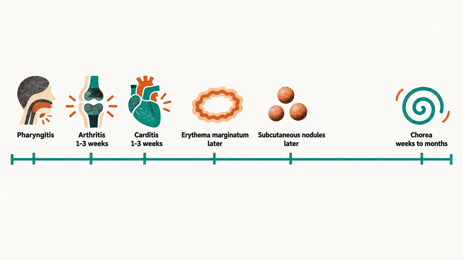
The five major manifestations — arthritis, carditis, erythema marginatum, subcutaneous nodules and — do not arrive together. They appear at different times after the infection and with very different frequencies. Carditis is the one that changes the long-term prognosis; arthritis is the one that brings the child to medical attention.
| Manifestation | Frequency | Timing after pharyngitis |
|---|---|---|
| Arthritis | The most common major manifestation in most series | Early — about 1-3 weeks |
| Carditis | Roughly half of cases, more when echocardiography is used systematically | Early — about 1-3 weeks |
| Erythema marginatum | Uncommon, under 10% | Later |
| Subcutaneous nodules | Uncommon, under 10% | Later |
| Sydenham’s chorea | Rare | Weeks to months |
Arthritis
Rheumatic arthritis has a recognisable signature:
- It affects large joints — knee, ankle, elbow, wrist — and rarely the small joints of the hands and feet.
- It is migratory, transient and asymmetric: one knee is inflamed for a few days and settles as another joint becomes involved.
- The classic signs of inflammation are present: pain, swelling, heat, redness and loss of function.
- The latency after pharyngitis is typically 2-3 weeks, and the arthritis responds dramatically to anti-inflammatory drugs.
- Monoarthritis does not exclude the diagnosis, particularly in populations at moderate or high risk.
When a child presents with joint inflammation after a sore throat, the main alternative diagnoses each differ from rheumatic arthritis in a way that can be checked:
- Juvenile idiopathic arthritis — persistent rather than migratory, and involving small joints symmetrically when it is the polyarticular form.
- Post-infectious reactive arthritis — develops during rather than after the infection, with normal inflammatory markers in many cases.
- Septic arthritis — a single hot joint that requires aspiration.
- Tuberculous arthritis — slower and monoarticular.
- Lyme disease — a history of tick exposure.
Carditis
Carditis is the manifestation that determines whether the child will need heart surgery or lifelong prophylaxis. Its spectrum is wide:
- Pancarditis — inflammation of endocardium, myocardium and pericardium together. In most cases the endocardium is the primary site, and the mitral valve is most commonly affected, followed by the aortic valve.
- A new murmur of mitral or aortic regurgitation is the classic clinical finding. Regurgitation is far more typical than stenosis, which belongs to the late, chronic phase.
- Echocardiography detects valvular regurgitation even when no murmur is audible. The 2015 revision of the diagnostic criteria accepts subclinical carditis — echocardiographic valvular regurgitation without an audible murmur — as a major manifestation, and Doppler echocardiography is recommended in every child with suspected rheumatic fever.
- First-degree atrioventricular block (a prolonged PR interval on the electrocardiogram) is the most common electrocardiographic finding; second- and third-degree block occur but are much less common.
- Severe carditis can present with heart failure and cardiomegaly, and it may be fatal during the acute episode, although death in the acute phase is rare overall.
Erythema marginatum
A non-pruritic, barely raised rash of reddish rings with a pale centre, 1-2 cm across, on the trunk and proximal limbs, often described as evanescent. The differential is the migratory erythema of early Lyme disease, and the way to separate them is the history of tick exposure.
Subcutaneous nodules
Firm, painless nodules 0.5-2 cm across, lying over tendons and bony prominences, which move with the skin and resolve spontaneously. They also occur in juvenile idiopathic arthritis and systemic lupus erythematosus, so they are not specific on their own.
Sydenham’s chorea
Chorea — involuntary, purposeless movement — is the rarest and the most difficult manifestation to recognise. It appears weeks to months after the streptococcal infection, often so long afterwards that the sore throat has been forgotten.
- Behavioural change — irritability, emotional lability, agitation, difficulty concentrating.
- Motor signs — uncoordinated, purposeless, involuntary movements, especially of the hands and face; deteriorating handwriting; facial grimacing.
- Movements disappear during sleep and are worsened by emotional stress.
- It is more common in girls.
- Echocardiography is often revealing, because most children with chorea have valvular involvement even when no murmur is heard. Unexplained chorea in a school-age girl, with mitral regurgitation on echocardiography, is a strong pointer to rheumatic fever.
Because the latency is so long, serological evidence of a preceding infection may have disappeared by the time chorea appears; a normal (the antibody test for a recent streptococcal infection) does not argue against the diagnosis in this situation.
Prognosis
The acute episode is usually survivable, and the arthritis and rash resolve without permanent damage. The prognosis is decided by the heart. Valve regurgitation may resolve, or may progress to scarring and stenosis over years to decades, with heart failure, atrial fibrillation and infective endocarditis as later complications.
Each recurrence of rheumatic fever adds further valve damage, so what happens after the first attack matters as much as the attack itself. Regular cardiology follow-up, adherence to prophylaxis — long-term antibiotics that protect the child from further streptococcal infection — and dental hygiene all matter for that reason.
Recognising the disease during the acute episode is what makes treatment, and later prophylaxis, possible. The diagnosis rests on criteria applied to a clinical picture rather than on any single test, and it is the balance of manifestations — not the severity of the sore throat — that identifies the child.