
Chronic anterior uveitis — inflammation of the iris and ciliary body at the front of the eye — is the complication of juvenile idiopathic arthritis (JIA) that justifies the most attention, because it usually produces none of the symptoms that would bring a child to a doctor. In most children there is no redness, no pain and no change in vision in the early stages, and the child appears entirely well. The eye is therefore looked at on a schedule rather than because of a complaint, and the examination that finds it is slit-lamp examination, a magnified examination with a narrow beam of light that can show inflammatory cells in the anterior chamber (the fluid-filled space behind the cornea) before anything is visible from the outside.
JIA is a group of chronic arthritides of childhood — arthritis beginning before the 16th birthday and lasting more than 6 weeks without a known cause — divided into categories by the number of joints involved and associated features. The risk of uveitis is not spread evenly across those categories: it follows particular categories and particular antibodies. That is what makes screening a matter of counting risk factors rather than of watching for symptoms.
What the untreated eye loses
Untreated chronic inflammation in the anterior chamber leaves permanent structural damage. — adhesions — form between the iris and the lens; cataract develops, sometimes accelerated by the corticosteroid drops used to treat the inflammation; glaucoma follows from both the inflammation and the treatment; and the end result can be permanent visual loss. Uveitis can also be present and active while the joints are quiet, so a child whose arthritis is well controlled may still have active eye disease. This is the reason a child with JIA needs a rheumatology and ophthalmology follow-up plan, and not only a prescription.
Who is screened, and how often
The frequency of screening is set by how many risk factors a child has. The risk factors are age at onset, antinuclear antibody (ANA) status — autoantibodies against components of the cell nucleus — the category of JIA, and how long the disease has lasted.
Children are at high risk when they have oligoarthritis, rheumatoid-factor-negative polyarthritis, psoriatic arthritis or undifferentiated arthritis, together with all three of: ANA positivity, onset before 7 years of age, and disease duration of 4 years or less. In current guidance these children are screened every 3 months.
Children are at lower risk when they have one of those same categories but are ANA negative, were 7 years or older at onset, or have had the disease for more than 4 years; this lower-risk group also includes and rheumatoid-factor-positive polyarthritis. These children are screened every 6-12 months, depending on the combination of risk factors they carry. Children with (arthritis with inflammation at tendon insertions), and those carrying HLA-B27, an inherited tissue-type marker, need screening as well, because they are at risk of both chronic anterior uveitis and the acute anterior uveitis of spondyloarthritis, the inflammatory arthritis of the spine and sacroiliac joints.
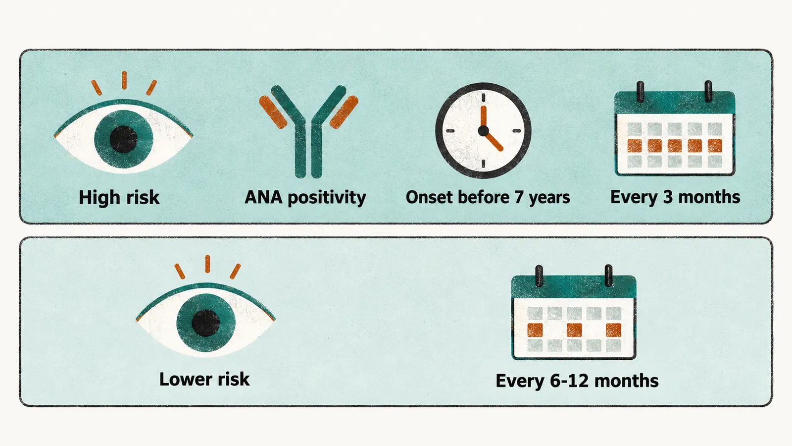
The schedule is adjusted by the ophthalmologist as risk changes, screening is continued for years, and it does not stop because the joints are quiet.
Once uveitis is found: treatment and monitoring
Treatment is stepped, and the aim is to control the inflammation while reducing exposure to topical steroids, which carry the risks of cataract and glaucoma:
- Topical glucocorticoids (corticosteroid eye drops) are the initial treatment. Prednisolone acetate 1% drops are the usual choice, and adding or increasing topical drops for short-term control is preferred to adding systemic glucocorticoids.
- When the eye still needs 1-2 drops a day of a topical glucocorticoid for control, systemic treatment is added or escalated so that the drops can be tapered. Subcutaneous methotrexate, the standard disease-modifying drug in JIA, is preferred to oral methotrexate for this purpose.
- When a biologic — an engineered protein that blocks one immune signal — is needed, the monoclonal antibody inhibitors of tumour necrosis factor (TNF), adalimumab and infliximab, are preferred to etanercept. In severe, sight-threatening uveitis, methotrexate and a monoclonal antibody TNF inhibitor are started together rather than methotrexate alone.
- When the eye disease is controlled on systemic treatment alone, therapy is not tapered until it has been well controlled for at least 2 years.
A child with established uveitis is monitored more closely than a child being screened, and the timing of examinations follows the treatment. While the disease is controlled on stable treatment, ophthalmic monitoring is no less frequently than every 3 months; when topical glucocorticoids are being tapered or stopped, monitoring follows within 1 month of each change; and when systemic treatment is changed, monitoring follows within 2 months of the change.
Uveitis affects the categories with an early, ANA-positive, oligoarticular pattern most, and it is uncommon in systemic JIA, which sits in the lower-risk group alongside rheumatoid-factor-positive polyarthritis. The danger in systemic JIA lies somewhere else entirely: in the systemic inflammation itself, and in the complication that can turn a controlled disease into an emergency within days.