Kawasaki disease is an acute, self-limiting vasculitis of medium-sized arteries — inflammation of the wall of arteries of that calibre — that occurs almost entirely in young children. Its importance comes from one target: the coronary arteries. Untreated, roughly a quarter of children develop coronary artery dilation or aneurysm (a localised ballooning of the artery), and those aneurysms can thrombose years later. Treated with (IVIG) inside the correct time window, the great majority recover with normal coronary arteries.
The diagnosis is clinical. There is no confirmatory laboratory test, and the fever is typically resistant to both antipyretics and antibiotics, so the diagnosis is usually reached only after a course of antibiotics has failed. Recognising it therefore starts with knowing which children are at risk.
Epidemiology
Kawasaki disease is a disease of the first years of life. Peak incidence is between 9 and 11 months of age; about 80% of patients are under 5 years old, and roughly half are under 2. Boys are affected somewhat more often than girls.
The incidence varies enormously with geography and ancestry. Japan has the highest reported rate in the world — approximately 359 per 100,000 children aged 0-4 in 2018 — followed by Korea and Taiwan. In the continental United States the figure is approximately 9 to 20 per 100,000 children under 5. Children of East Asian ancestry have a higher risk wherever they live, which points to a genetic contribution rather than a purely environmental one.
The timing of cases adds an environmental clue. Cases cluster in winter and spring in temperate climates, and outbreaks occur. A respiratory or other febrile illness in the two to three weeks before the onset of the classic features is commonly reported. Together these patterns suggest an infectious trigger acting in a genetically susceptible child, but no single organism has been identified, and the disease is not transmitted in any simple person-to-person way.
The clinical features
The fever of Kawasaki disease is high, sustained, and poorly responsive to antipyretics and to antibiotics. It lasts at least 5 days if untreated, and most children have had a course of antibiotics for a presumed bacterial infection before the diagnosis is made.
Five principal features accompany the fever.
| Feature | What to look for |
|---|---|
| Bilateral | Red eyes without exudate — no pus, no crusting, no matting of the lashes. Unilateral or exudative conjunctivitis argues against the diagnosis |
| Oral cavity changes | Dry, cracked, fissured lips, sometimes with blood crusts; strawberry tongue (reddened papillae through a desquamating white coating); hyperaemic pharynx without vesicles or tonsillar exudate |
| Polymorphous rash | A pink maculopapular exanthem (widespread rash) of trunk and limbs, not specific to the disease and easily mistaken for a viral rash |
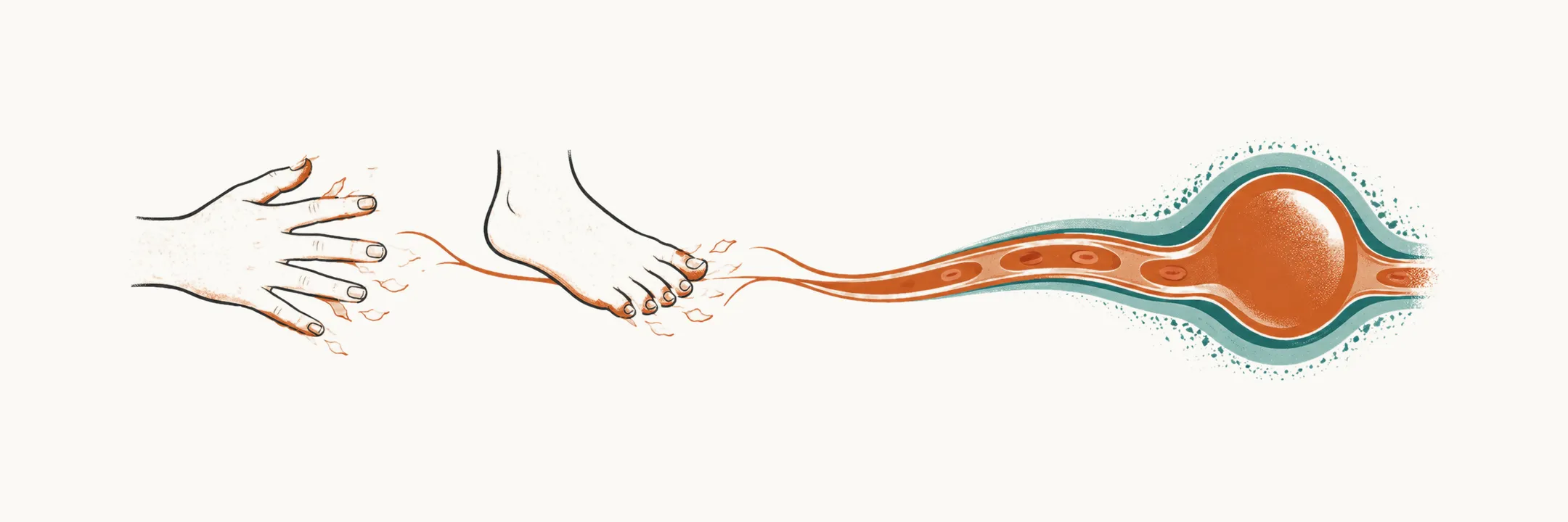
| Extremity changes | Erythema and oedema of the hands and feet, often more prominent on the feet, during the febrile phase. Periungual desquamation (peeling of the skin around the nails) of the fingers and toes appears 2-3 weeks later |
| Cervical lymphadenopathy | Usually unilateral, one node or one side of the neck. Bilateral or generalised lymphadenopathy suggests another diagnosis |
Periungual desquamation is highly suggestive of Kawasaki disease but appears too late to count towards the diagnostic criteria; it is most useful retrospectively, in a child whose fever has already settled.
Other findings are common but do not count towards the criteria: marked irritability, abdominal pain, vomiting or diarrhoea, arthralgia or arthritis, sterile pyuria (white cells in the urine without bacterial growth), aseptic meningitis, and hydrops (distension) of the gallbladder.
The timing of the findings
The features do not all appear at once. The illness moves through three phases:
| Phase | Time from fever onset | Typical findings |
|---|---|---|
| Acute | Days 1-7 | Fever and the five principal features; irritability; myocardial and pericardial inflammation (often subclinical) |
| Subacute | Weeks 2-3 | Coronary artery dilation and aneurysm; periungual desquamation; rising platelet count |
| Convalescent | Week 4 onwards | Clinical features resolve; risk of coronary thrombosis if an aneurysm has formed; risk stratification guided by cardiac involvement |
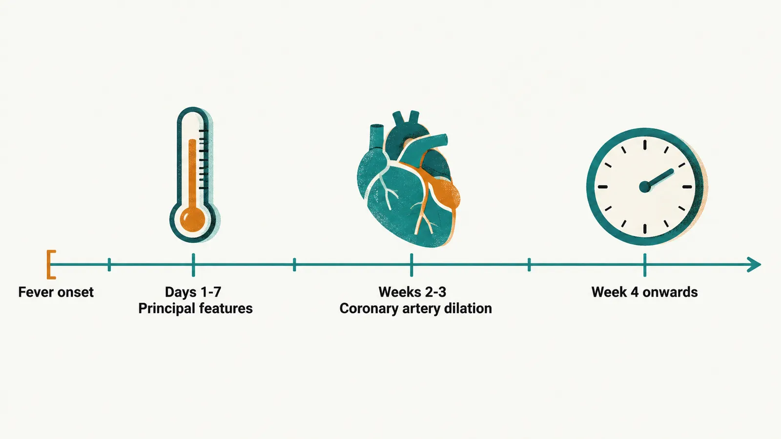
The three phases explain why the illness is easy to miss at the first consultation. The features that are most specific come later, and the finding that matters most — coronary artery dilation — appears in the second and third week, after the point at which treatment is most effective.
Differential diagnosis
Because the features are individually non-specific and the most specific ones come late, the differential is broad. Most of it is infection:
- Epstein-Barr virus and other viral infections — fever, rash and lymphadenopathy. Atypical lymphocytes, pharyngitis with exudate and splenomegaly point away from Kawasaki disease.
- Scarlet fever — shares the , but the rash is fine, sandpaper-like, and the child has pharyngitis with a positive streptococcal test.
- Measles, roseola infantum and other exanthems — usually separated by the absence of conjunctival, oral and extremity changes.
- A bacterial infection that has failed to respond to antibiotics, including urinary tract infection when there is sterile pyuria and pneumonia when there are respiratory findings.
Two inflammatory illnesses share several of its features, and a third alternative is identified from the history:
- (systemic JIA) — the form of childhood arthritis dominated by fever and inflammation outside the joints. It shares fever, rash and arthritis with Kawasaki disease. The fever pattern differs (daily spikes in systemic JIA, sustained fever in Kawasaki disease) and the echocardiogram is decisive, showing coronary involvement in Kawasaki disease and, when present at all, pericardial involvement in systemic JIA.
- Multisystem inflammatory syndrome following SARS-CoV-2 infection — a distinct post-infectious hyperinflammatory illness that can also produce fever, rash, conjunctivitis and coronary involvement, and which shares features with Kawasaki disease. It is treated differently, and it is distinguished by age, the epidemiological link to a preceding infection, and a different pattern of organ involvement.
- Drug reactions and hypersensitivity — the history is the discriminator.
No single feature identifies the disease, and the diagnosis is made from how many of the principal features are present, how long the fever has lasted, and what the laboratory tests and the echocardiogram show.