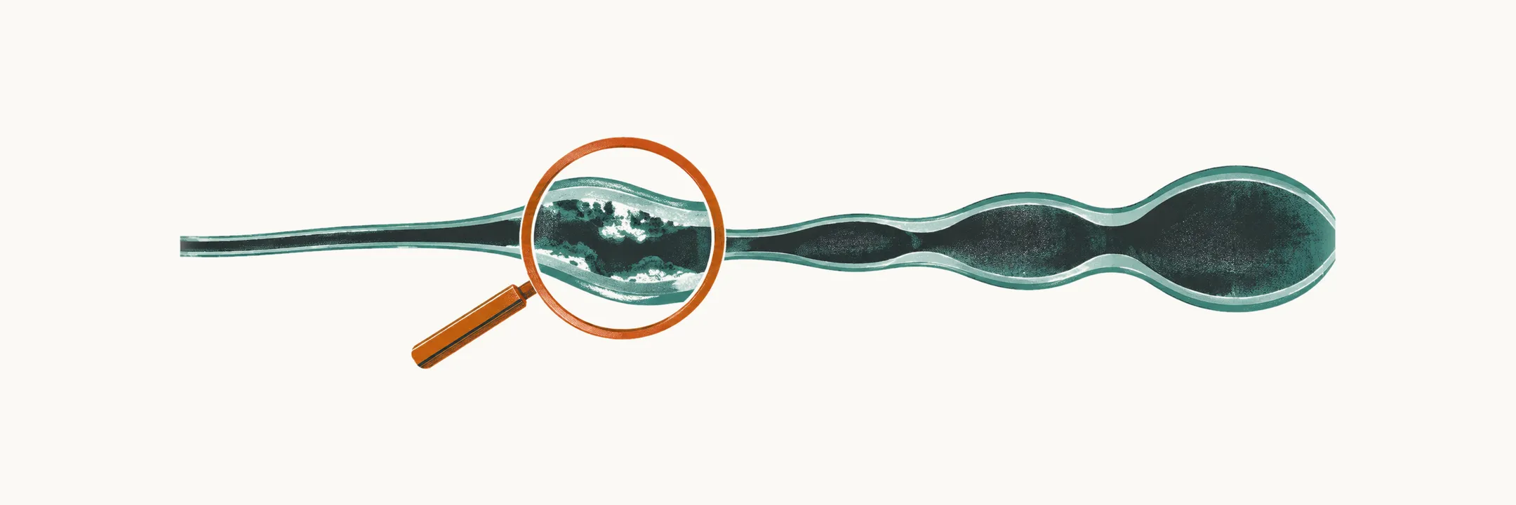
Kawasaki disease is an acute vasculitis — inflammation of the vessel wall — of medium-sized arteries that occurs almost entirely in young children, and its danger lies in the damage it can do to the coronary arteries. It is diagnosed clinically: there is no confirmatory laboratory test. Because the fever is typically resistant to both antipyretics and antibiotics, the diagnosis is usually reached only after a course of antibiotics for a presumed bacterial infection has failed.
The five principal features that count towards the diagnosis are bilateral without exudate, changes of the lips and oral cavity, a polymorphous rash (one that can take several forms), erythema and oedema of the hands and feet, and cervical lymphadenopathy — usually unilateral. Findings such as marked irritability, arthralgia, hydrops of the gallbladder and sterile pyuria are common in Kawasaki disease but do not count towards the rule.
The diagnostic rule
Fever for at least 5 days plus at least 4 of the 5 principal features makes the diagnosis of complete (classical) Kawasaki disease. When 4 or more features are present, the diagnosis can be made from day 4 of fever, so waiting for the fifth day is not necessary.
Fever for at least 5 days plus 2 or 3 features, together with supportive laboratory findings or echocardiographic coronary changes, is generally called . It is the usual presentation in infants under 1 year, who are the group most likely to be missed.
Terminology varies between sources. Some use “incomplete” and “atypical” interchangeably for any presentation that does not meet the full criteria, while others reserve atypical for the child whose findings are unusual rather than merely fewer — pericarditis, myocarditis, aseptic meningitis, arthritis, severe gastrointestinal involvement, or respiratory findings. Both describe the same disease; the practical point is that a child can have Kawasaki disease without ever meeting the classical rule. For those children, the laboratory tests and the echocardiogram carry the weight of the diagnosis.
Laboratory findings
Nothing on this list confirms the diagnosis. The tests reflect the systemic inflammation of the acute illness; they support the diagnosis and drive the incomplete-disease algorithm. Two general markers of inflammation appear throughout: C-reactive protein (CRP) and the erythrocyte sedimentation rate (ESR).
| Test | Pattern in Kawasaki disease |
|---|---|
| White blood cells | Raised with neutrophil predominance |
| Haemoglobin | Mild anaemia in the acute phase; early anaemia is associated with more severe disease |
| Platelets | Normal early, rising in the subacute phase — too late to help the diagnosis |
| CRP and ESR | Both raised; ESR falls more slowly than CRP |
| Alanine aminotransferase (ALT) | Mildly raised |
| Albumin | Low |
| Urinalysis | Sterile pyuria — useful for distinguishing the febrile child from urinary tract infection |
The incomplete disease algorithm
The widely used algorithm for the child with fever and fewer than 4 features starts from CRP and ESR, and then counts the supportive laboratory findings:
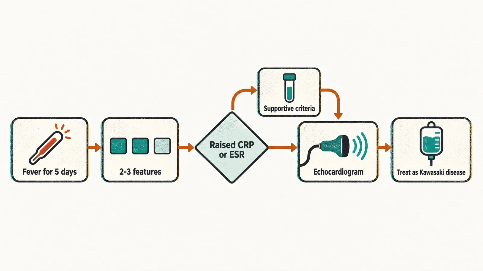
- Fever for 5 days or more with 2-3 features, and normal CRP and ESR: continue clinical and laboratory monitoring with serial echocardiography. If coronary abnormalities appear, the diagnosis is made.
- Fever for 5 days or more with 2-3 features, and raised CRP or ESR: obtain further laboratory tests — full blood count, liver enzymes, albumin, urinalysis. If 3 or more of 6 supportive criteria are present (anaemia for age, platelet count 450,000/mm³ or more after day 7, albumin 3.0 g/dL or less, raised ALT, white cell count 15,000/mm³ or more, 10 or more white cells per high-power field on urinalysis), obtain an echocardiogram and treat as Kawasaki disease.
- Coronary abnormalities on echocardiography at any point establish the diagnosis regardless of the laboratory results.
Infants are handled separately because they present with fewer features. An infant 6 months old or younger with fever for 7 days or more and no other explanation should have laboratory testing, and an echocardiogram if there is evidence of systemic inflammation, even with no principal features at all.
Echocardiography
Echocardiography is the most important investigation, and the only one that can establish the diagnosis in the incomplete form:
- At diagnosis, to look for early coronary involvement. A normal study at this stage does not exclude the disease, because aneurysms form later.
- During follow-up, because the coronary arteries must be re-examined as the disease evolves. Repeat studies are typically done at diagnosis and again at about 1-2 weeks and 4-6 weeks.
- For risk stratification, because the severity of coronary involvement determines the long-term plan.
Coronary artery dimensions in children are reported as Z-scores — standard deviations from the expected value for body surface area — because absolute diameters are uninterpretable across ages and sizes. The usual grading is: dilation at a Z-score of 2 to less than 2.5; a small aneurysm at 2.5 to less than 5; a medium aneurysm at 5 to less than 10; and a giant aneurysm at 10 or more.
Because the coronary arteries can be normal on the day the diagnosis is made and abnormal two weeks later, echocardiography establishes which children already have coronary involvement. Preventing that involvement is a separate decision, taken on clinical grounds before any of these results are available.