
Graves disease is the most common cause of thyrotoxicosis, accounting for about 60-80% of cases. It is roughly ten times more common in women than in men: the prevalence is about 2% in women and 0.2% in men. Whether it develops depends on a person’s genetic predisposition together with their level of exposure to iodine.
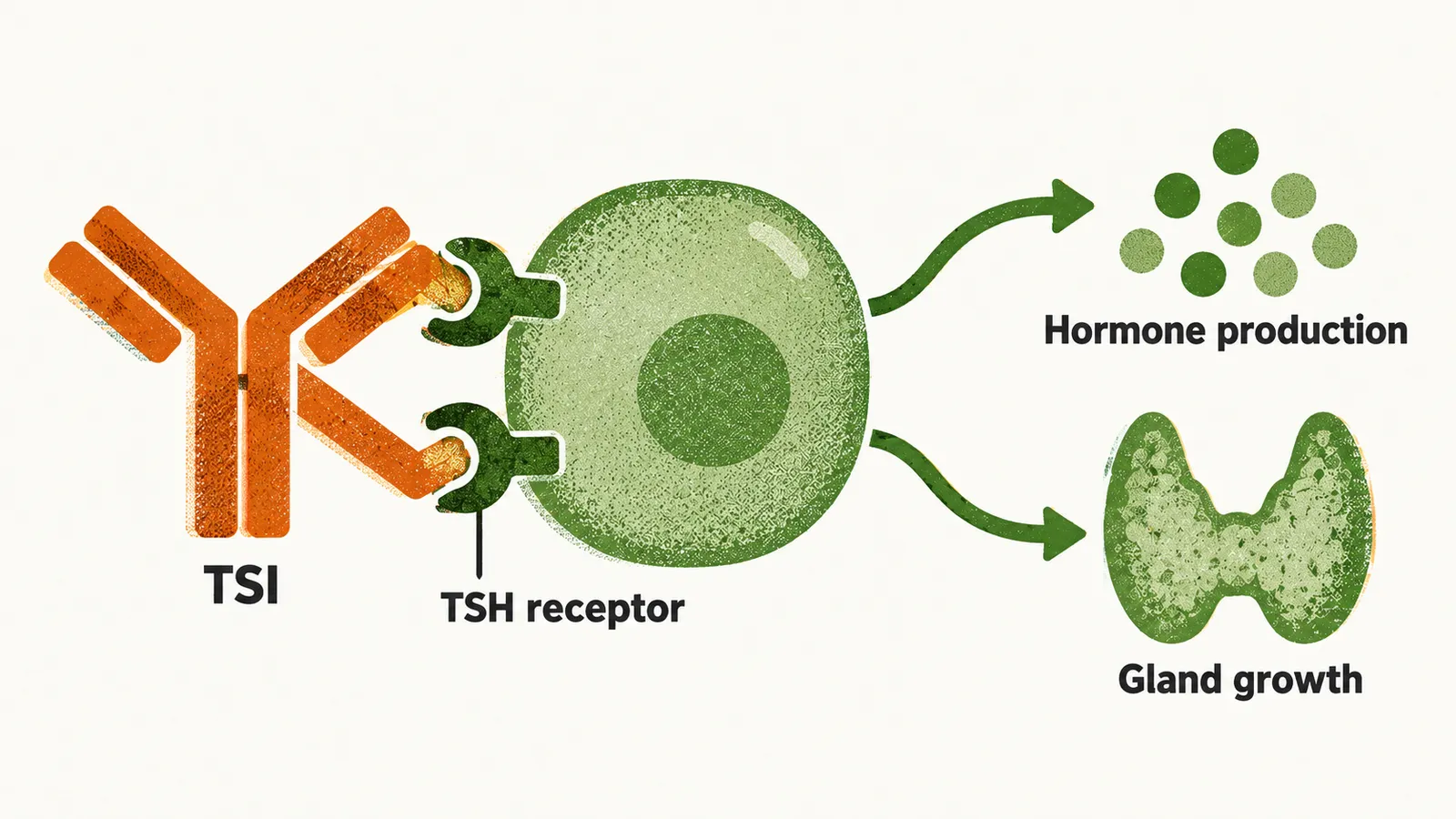
The hyperthyroidism of Graves disease is caused by the thyroid-stimulating immunoglobulins (TSIs), antibodies made by lymphocytes that reside in the thyroid gland, the bone marrow and the lymph nodes. They bind the TSH receptor on the thyroid cell (the receptor for TSH, the pituitary thyroid-stimulating hormone) and stimulate it in place of TSH, so hormone production and gland growth proceed without feedback control. The combination of thyrotoxicosis with TSH-receptor antibodies in the blood is what defines Graves disease.
Antibodies against thyroid peroxidase (TPO, the enzyme that builds thyroid hormone) and against thyroglobulin (Tg, the protein in which the hormone is made and stored) are also present in more than 80% of cases. Because a thyroiditis can damage the gland at the same time as the TSIs stimulate it, and because the balance between the two differs between patients, the clinical progression of Graves disease is difficult to predict.
What sets the autoimmunity going is a mix of inherited risk and environmental triggers. As in autoimmune hypothyroidism (Hashimoto thyroiditis), inherited variation predisposes to Graves disease. The main genes identified are:
- HLA-DR polymorphisms
- CTLA-4 polymorphism
- CD25 polymorphism
- CD226 polymorphism
- PTPN22 polymorphism
- a TSH-R polymorphism
HLA-DR variants change how thyroid antigens are presented to T cells, while CTLA-4, CD25, CD226 and PTPN22 regulate the signalling that keeps T cells in check, and the TSH-R polymorphism alters the receptor the antibodies target. Together they lower the threshold at which the immune system attacks the gland.
On this background, a sudden increase in iodine intake raises the incidence of Graves disease, and the incidence rises three-fold after childbirth. Some drugs that disturb immune regulation can also bring it on: highly active antiretroviral therapy (HAART), immune checkpoint inhibitors and alemtuzumab.
The same immune attack also reaches the eye, because the TSH receptor is expressed in the orbit as well. Ophthalmopathy is one of the most prominent clinical manifestations of thyrotoxicosis, including Graves disease. It begins when T cells infiltrate the extraocular muscles and release cytokines such as TNF, IL-1 and IFN. These cytokines activate orbital fibroblasts to make glycosaminoglycans (GAGs), which are hydrophilic and trap water in the tissues around and behind the eyeball. In the late stage the process turns into irreversible fibrosis with fat accumulation around the eye, and this can cause diplopia, proptosis and optic neuropathy.