
Adipose tissue is an active participant in type 2 diabetes mellitus, or T2DM, rather than a passive fat store. Insulin acts on adipocytes in two ways that matter for lipid metabolism: it inhibits lipolysis, the breakdown of stored triglyceride, and it drives the uptake of free fatty acids (FFA) from the plasma into the cells.
In patients with T2DM the adipocytes become insulin-resistant, so both effects weaken. FFA are no longer taken up from the plasma, which produces a day-long high concentration of circulating FFA. The excess FFA then work against insulin themselves, worsening the sensitivity of tissues, and they accumulate in the liver and muscle, where they cause lipotoxicity, direct injury from lipid overload. More than 85% of the patients in the US who have T2DM are obese, which places adipose tissue at the centre of the disease.
Adipokines and inflammation
The presence of chronic, low-grade inflammation of the adipocytes has been suggested to increase the chance of insulin resistance and of developing T2DM. That inflammation is largely commanded by cytokines released by the adipocytes themselves, known as adipokines. Several adipokines are relevant to the pathogenesis of T2DM:
- Adiponectin is inversely related to the metabolic syndrome, T2DM and atherosclerosis. It increases the oxidation of FFA and reduces glucose production by the liver, and it is anti-inflammatory, suppressing the effect of tumour necrosis factor (TNF) and NF-κB. Its fall in obesity removes a protective signal.
- Human resistin is a cytokine released by the immune cells that infiltrate adipose tissue, triggering those cells to produce adipokines.
- Leptin is physiologically a suppressor of appetite. Obese patients are resistant to it, so hyperleptinemia can be found in them, and hyperleptinemia is associated with an inflammatory state in the body.
- Visfatin is an adipokine that is increased in obese patients and has a pro-inflammatory effect.
How free fatty acids promote insulin resistance
A high level of FFA in the cells and in the circulation promotes insulin resistance through several mechanisms: an oxidative burden inside the cell with subsequent endoplasmic reticulum stress, inhibition of the insulin receptor substrate (IRS), the docking protein that carries the insulin signal inside the cell, and a reduction of insulin’s vasodilatory effect on the endothelium, because nitric oxide (NO) production is controlled by the IRS pathway.
The Randle cycle explains the effect of accumulated FFA on glucose use. A high FFA level drives high β-oxidation of FFA in the mitochondria, and that high β-oxidation chain-reacts through the cell. It produces a large amount of acetyl-CoA, which inhibits pyruvate dehydrogenase, and it raises the NADH/NAD ratio, which slows the Krebs cycle because its enzymes are NADH-dependent. The stalled cycle lets citrate accumulate, and citrate is a potent inhibitor of the enzyme PFK, so glucose-6-phosphate accumulates too and the entry of glucose into the muscle cell falls. With less glucose entering, there is less glycogen synthesis and less glucose oxidation.
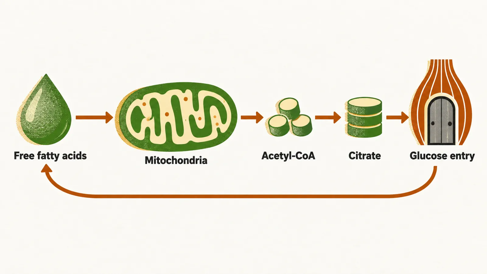
The conclusion from the Randle cycle is that a high oxidation rate of FFA reduces the uptake, oxidation and use of glucose in the cells. More recent studies in humans have added that a rise in FFA β-oxidation does not only reduce glucose oxidation and glycogen formation: it can also induce insulin resistance through other signalling pathways, inhibiting PKC, which inhibits IRS, and inhibiting PKB, which inhibits glycogen synthase.
From fatty liver to fibrosis
With the rising incidence of metabolic dysfunction-associated steatotic liver disease (MASLD), formerly called non-alcoholic fatty liver disease, complications tightly related to insulin resistance in T2DM have become more common. In MASLD there is steatosis, the accumulation of FFA inside the hepatocytes, which makes them vulnerable to injury and fibrosis. In patients with T2DM the same progressive insulin resistance reaches the hepatocytes, and the accumulating FFA lead to MASLD formation and to progression toward its severe and fibrotic forms. That progression is driven by two processes acting together: insulin resistance with further FFA accumulation in the hepatocytes, and the formation of reactive oxygen species during the β-oxidation of those FFA.