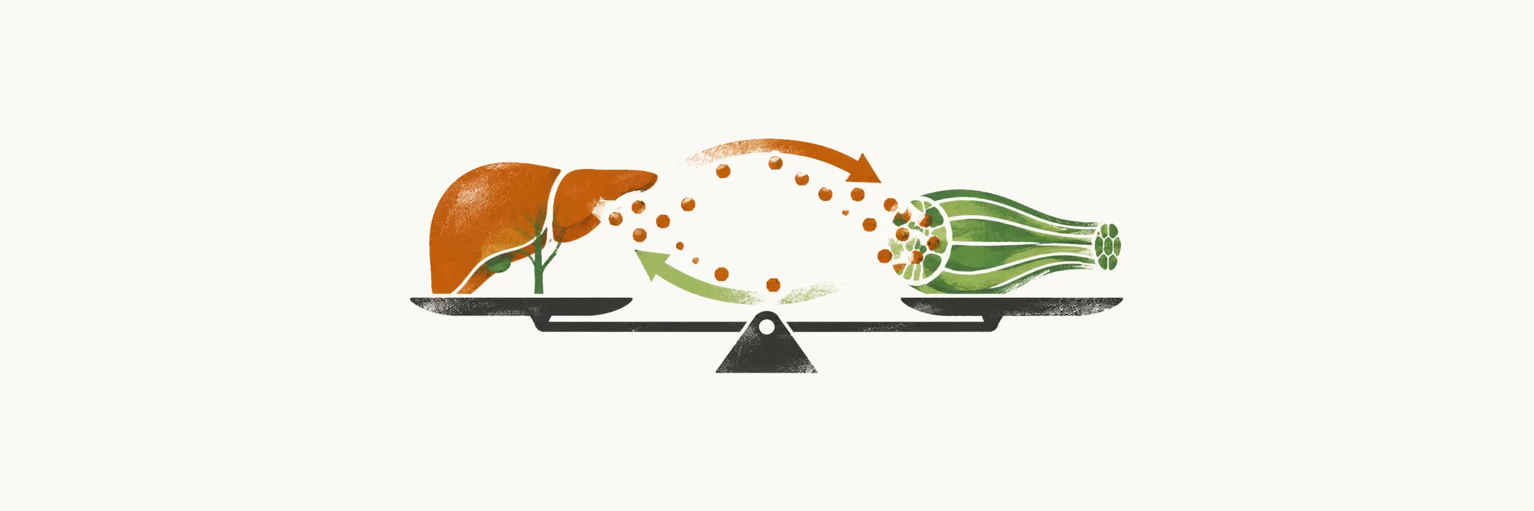
Normal glucose homeostasis keeps blood glucose steady between meals and disposes of a glucose load after eating. It depends on a match between the glucose the body produces and the glucose its tissues take up, and on the hyperbolic relation between insulin sensitivity and β-cell function: the product of the two stays roughly constant, so the higher the insulin sensitivity, the lower the β-cell function needed, and vice versa. A change in one therefore shifts the other.
The state of glucose in the body is described in two phases:
- post-absorptive state — 10–12 hours after fasting
- post-prandial state — after ingestion of glucose
In the post-absorptive state the basal glucose utilisation rate averages around 2 mg/kg.min, which precisely matches endogenous glucose production (EGP) — the glucose the body itself releases into the blood. More than 85% of this production comes from the liver, with a smaller contribution from the kidney.
Post-absorptive state
In the post-absorptive state, the glucose that is used in the body is distributed among three consumers:
- brain — 50%, which is insulin-independent
- splanchnic area (liver plus gastrointestinal tract) — 25%, which is insulin-independent
- insulin-dependent tissue, mainly muscle tissue — 25%
Post-prandial state
During the post-absorptive state there is a perfect balance between endogenous glucose production and the body’s need for glucose, but that balance is disrupted after the introduction of exogenous glucose. After a meal containing glucose, the resulting hyperglycemia and hyperinsulinemia increase the absorption of glucose into splanchnic and peripheral tissues — of the glucose taken up in the periphery, about 85% is disposed of in muscle tissue and 5% is metabolised by adipocytes. At the same time, insulin reduces endogenous glucose production and lowers the concentration of free fatty acids (FFA) in the plasma.
The very first and rate-limiting step in insulin’s effect on peripheral tissues, mainly muscle tissue, is the transport of insulin from plasma to the muscle cells. This step depends mainly on the insulin receptors on the endothelial cells and on the activation of nitric oxide (NO) synthase in those cells.
In normal subjects, the rate of whole-body glucose uptake when glucose is introduced through the intravenous route is measured at around 7 mg/kg.min. Three points about that number matter:
- the glucose uptake by the CNS stays constant, at around 1–1.2 mg/kg.min, in both normal and diabetic patients and in both the post-absorptive and post-prandial states, because it is insulin-independent
- the glucose uptake by the splanchnic areas, mainly the liver, in the post-prandial state is only about 0.5 mg/kg.min, similar in both normal subjects and patients with type 2 diabetes
- the majority of the glucose is taken up by muscle tissue, more than 85%
These results relate to the intravenous route. Oral ingestion of glucose distributes the load differently:
- splanchnic tissues take up 28% of the oral glucose
- peripheral tissues take up 72% of the oral glucose
- within that peripheral fraction, brain uptake is still 1–1.2 mg/kg.min, and muscle tissue takes up 45% of the total oral glucose
- basal hepatic glucose production (HGP) declines by 53%, so after oral glucose the endogenous glucose production (EGP) is less suppressed than after intravenous glucose
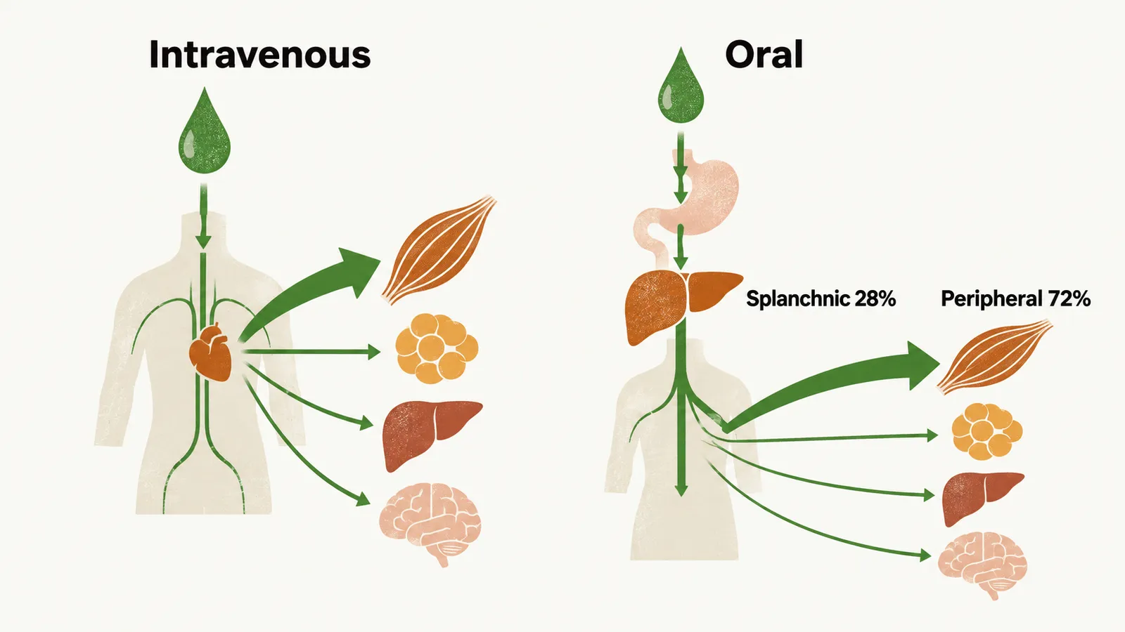
Three differences between the two routes follow. Splanchnic uptake plays an important role after oral glucose, because the gut and liver see the glucose before the rest of the body does. Peripheral tissue uptake is quantitatively smaller by this route, though it is still the largest single destination. And endogenous glucose continues to be produced after oral ingestion, because the suppression of EGP is incomplete.