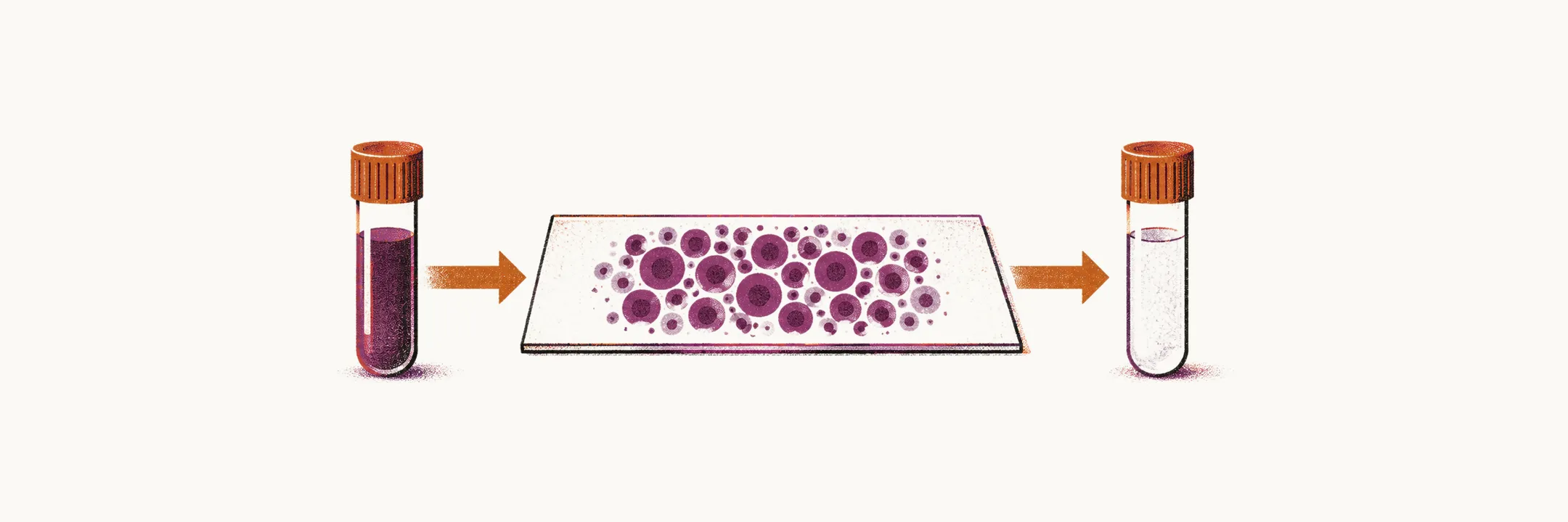
The diagnosis of acute lymphoblastic leukemia (ALL) starts with the peripheral blood and the bone marrow: the blood findings raise the suspicion, the bone marrow aspirate confirms the leukemia, and the lumbar puncture detects central nervous system involvement.
Peripheral blood and bone marrow
The very first and main steps for the diagnosis of ALL are the testing of peripheral blood and bone marrow.
Peripheral blood counts in ALL are very variable, so the peripheral blood white cell count most of the time cannot be used as a diagnostic tool on its own. Characteristically the blood shows anemia, thrombocytopenia and neutropenia, but the details differ widely. The white cell count is normal or reduced in 40% of cases, and is above 100,000 per microliter in only 16% of cases; no blasts are found in the peripheral blood in 8% of cases. About one-third of patients have a hemoglobin level below 7–8 g/dL, and a platelet count below 20,000 per microliter occurs in one-fifth of adults with ALL.
Because the blood cannot settle the question, a bone marrow aspirate (a sample of marrow drawn through a needle) is essential for confirming the leukemia and distinguishing between acute myeloid leukemia (AML) and ALL.
The bone marrow is usually heavily packed with blast cells, which make up about 90% of the bone marrow in about 70% of patients.
Differential diagnosis
Other conditions can resemble ALL before the marrow is examined. The differential diagnosis of ALL includes:
- infection
- chronic myeloid leukemia (CML) in acute blastic crisis
- non-Hodgkin lymphoma (NHL) with bone marrow involvement
Cerebrospinal fluid
Lumbar puncture, in which a needle samples the cerebrospinal fluid (CSF) that surrounds the brain and spinal cord, is a routine part of the diagnosis of ALL; when more than 5 morphologically confirmed blast cells per microliter of CSF are present, CNS leukemia is confirmed.
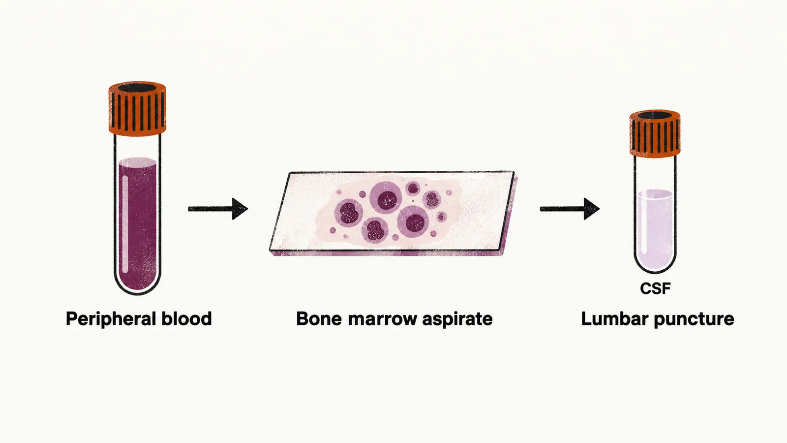
For prevention of possible transferred blast cells from the peripheral blood to the CSF at the first lumbar puncture, intrathecal methotrexate should be used.
Once the diagnosis is established, other techniques such as flow cytometry and cell staining are used for further classification of the leukemia.